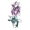
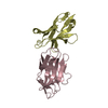
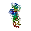
- PDB-4nfa: Structure of the C-terminal doamin of Knl1 -
+
Open data
ID or keywords:
Loading...
-
Basic information
Entry
Database: PDB / ID: 4nfa
Title
Structure of the C-terminal doamin of Knl1
Components
Protein CASC5
Keywords
CELL CYCLE / RWD domain
Function / homology
Function and homology information
Knl1/Spc105 complex / positive regulation of meiosis I spindle assembly checkpoint / homologous chromosome orientation in meiotic metaphase I / acrosome assembly / regulation of mitotic cell cycle spindle assembly checkpoint / attachment of spindle microtubules to kinetochore / outer kinetochore / protein localization to kinetochore / mitotic sister chromatid segregation / Amplification of signal from unattached kinetochores via a MAD2 inhibitory signal ...Knl1/Spc105 complex / positive regulation of meiosis I spindle assembly checkpoint / homologous chromosome orientation in meiotic metaphase I / acrosome assembly / regulation of mitotic cell cycle spindle assembly checkpoint / attachment of spindle microtubules to kinetochore / outer kinetochore / protein localization to kinetochore / mitotic sister chromatid segregation / Amplification of signal from unattached kinetochores via a MAD2 inhibitory signal / Mitotic Prometaphase / Deposition of new CENPA-containing nucleosomes at the centromere / EML4 and NUDC in mitotic spindle formation / acrosomal vesicle / Resolution of Sister Chromatid Cohesion / RHO GTPases Activate Formins / kinetochore / Separation of Sister Chromatids / microtubule binding / nuclear body / cell division / nucleoplasm / nucleus / cytosol Similarity search - Function
Journal: Mol Cell / Year: 2014 Title: Modular assembly of RWD domains on the Mis12 complex underlies outer kinetochore organization. Authors: Arsen Petrovic / Shyamal Mosalaganti / Jenny Keller / Marta Mattiuzzo / Katharina Overlack / Veronica Krenn / Anna De Antoni / Sabine Wohlgemuth / Valentina Cecatiello / Sebastiano ...Authors: Arsen Petrovic / Shyamal Mosalaganti / Jenny Keller / Marta Mattiuzzo / Katharina Overlack / Veronica Krenn / Anna De Antoni / Sabine Wohlgemuth / Valentina Cecatiello / Sebastiano Pasqualato / Stefan Raunser / Andrea Musacchio / Abstract: Faithful chromosome segregation is mandatory for cell and organismal viability. Kinetochores, large protein assemblies embedded in centromeric chromatin, establish a mechanical link between ...Faithful chromosome segregation is mandatory for cell and organismal viability. Kinetochores, large protein assemblies embedded in centromeric chromatin, establish a mechanical link between chromosomes and spindle microtubules. The KMN network, a conserved 10-subunit kinetochore complex, harbors the microtubule-binding interface. RWD domains in the KMN subunits Spc24 and Spc25 mediate kinetochore targeting of the microtubule-binding subunits by interacting with the Mis12 complex, a KMN subcomplex that tethers directly onto the underlying chromatin layer. Here, we show that Knl1, a KMN subunit involved in mitotic checkpoint signaling, also contains RWD domains that bind the Mis12 complex and that mediate kinetochore targeting of Knl1. By reporting the first 3D electron microscopy structure of the KMN network, we provide a comprehensive framework to interpret how interactions of RWD-containing proteins with the Mis12 complex shape KMN network topology. Our observations unveil a regular pattern in the construction of the outer kinetochore.
ProteinCASC5 / ALL1-fused gene from chromosome 15q14 protein / AF15q14 / Bub-linking kinetochore protein / Blinkin ...ALL1-fused gene from chromosome 15q14 protein / AF15q14 / Bub-linking kinetochore protein / Blinkin / Cancer susceptibility candidate gene 5 protein / Cancer/testis antigen 29 / CT29 / Kinetochore-null protein 1 / Protein D40/AF15q14
Mass: 24173.303 Da / Num. of mol.: 1 / Fragment: Knl1 C-terminal domain, UNP residues 2131-2337 Source method: isolated from a genetically manipulated source Source: (gene. exp.) Homo sapiens (human) / Gene: CASC5, KIAA1570, KNL1 / Production host: Escherichia coli (E. coli) / References: UniProt: Q8NG31
In the structure databanks used in Yorodumi, some data are registered as the other names, "COVID-19 virus" and "2019-nCoV". Here are the details of the virus and the list of structure data.
Jan 31, 2019. EMDB accession codes are about to change! (news from PDBe EMDB page)
EMDB accession codes are about to change! (news from PDBe EMDB page)
The allocation of 4 digits for EMDB accession codes will soon come to an end. Whilst these codes will remain in use, new EMDB accession codes will include an additional digit and will expand incrementally as the available range of codes is exhausted. The current 4-digit format prefixed with “EMD-” (i.e. EMD-XXXX) will advance to a 5-digit format (i.e. EMD-XXXXX), and so on. It is currently estimated that the 4-digit codes will be depleted around Spring 2019, at which point the 5-digit format will come into force.
The EM Navigator/Yorodumi systems omit the EMD- prefix.
Related info.:Q: What is EMD? / ID/Accession-code notation in Yorodumi/EM Navigator
Yorodumi is a browser for structure data from EMDB, PDB, SASBDB, etc.
This page is also the successor to EM Navigator detail page, and also detail information page/front-end page for Omokage search.
The word "yorodu" (or yorozu) is an old Japanese word meaning "ten thousand". "mi" (miru) is to see.
Related info.:EMDB / PDB / SASBDB / Comparison of 3 databanks / Yorodumi Search / Aug 31, 2016. New EM Navigator & Yorodumi / Yorodumi Papers / Jmol/JSmol / Function and homology information / Changes in new EM Navigator and Yorodumi
 Movie
Movie Controller


 Open data
 Basic information
 Components
 Keywords
 Function and homology information
 Homo sapiens (human)
 X-RAY DIFFRACTION /
  Authors
 Citation

 Structure visualization
 Downloads & links
 Other downloads


 PDBj
PDBj


 Assembly


 Mass: 35.453 Da / Num. of mol.: 1 / Source method: obtained synthetically / Formula: Cl
 Mass: 92.094 Da / Num. of mol.: 3 / Source method: obtained synthetically / Formula: C3H8O3
 Mass: 18.015 Da / Num. of mol.: 101 / Source method: isolated from a natural source / Formula: H2O
 Sample preparation
 / Beamline: ID14-1 / Wavelength: 0.933 Å
 Processing