 Movie
Movie Controller

[English] 日本語
 Yorodumi
- PDB-1ixv: Crystal Structure Analysis of homolog of oncoprotein gankyrin, an... -
+ Open data

- Basic information
| Entry | Database: PDB / ID: 1ixv | ||||||
|---|---|---|---|---|---|---|---|
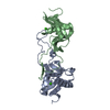
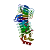
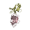
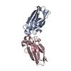
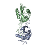
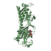
| Title | Crystal Structure Analysis of homolog of oncoprotein gankyrin, an interactor of Rb and CDK4/6 | ||||||
 Components Components | Probable 26S proteasome regulatory subunit p28 | ||||||
 Keywords Keywords | PROTEIN BINDING / Ankyrin repeats | ||||||
| Function / homology |  Function and homology information Function and homology informationproteasome regulatory particle binding / proteasome regulatory particle assembly / protein folding chaperone / nucleus / cytosol Similarity search - Function | ||||||
| Biological species |  | ||||||
| Method |  X-RAY DIFFRACTION / SYNCHROTRON / MIR / Resolution: 2.3 Å X-RAY DIFFRACTION / SYNCHROTRON / MIR / Resolution: 2.3 Å | ||||||
 Authors Authors | Padmanabhan, B. / Adachi, N. / Kataoka, K. / Horikoshi, M. | ||||||
 Citation Citation | Journal: J.BIOL.CHEM. / Year: 2004 Title: Crystal structure of the homolog of the oncoprotein gankyrin, an interactor of Rb and CDK4/6 Authors: Padmanabhan, B. / Adachi, N. / Kataoka, K. / Horikoshi, M. | ||||||
| History |
|
- Structure visualization
| Structure viewer | Molecule: MolmilJmol/JSmol |
|---|
- Downloads & links
-Download
| PDBx/mmCIF format | 1ixv.cif.gz | 57.7 KB | Display | PDBx/mmCIF format |
|---|---|---|---|---|
| PDB format | pdb1ixv.ent.gz | 42.2 KB | Display | PDB format |
| PDBx/mmJSON format | 1ixv.json.gz | Tree view | PDBx/mmJSON format | |
| Others |  Other downloads Other downloads |
-Validation report
| Arichive directory | https://data.pdbj.org/pub/pdb/validation_reports/ix/1ixvftp://data.pdbj.org/pub/pdb/validation_reports/ix/1ixv | HTTPS FTP |
|---|
-Related structure data
| Similar structure data |
|---|


-Links
 PDBj
PDBj

- Assembly
| Deposited unit | 
| ||||||||
|---|---|---|---|---|---|---|---|---|---|
| 1 |
| ||||||||
| Unit cell |
|
-Components
| #1: Protein | Mass: 25933.566 Da / Num. of mol.: 1 Source method: isolated from a genetically manipulated source Source: (gene. exp.) Plasmid: pET28b-Nas6p / Production host:  |
|---|---|
| #2: Water | ChemComp-HOH /  Mass: 18.015 Da / Num. of mol.: 102 / Source method: isolated from a natural source / Formula: H2O Mass: 18.015 Da / Num. of mol.: 102 / Source method: isolated from a natural source / Formula: H2O |
-Experimental details
-Experiment
| Experiment | Method: X-RAY DIFFRACTION / Number of used crystals: 1 |
|---|
- Sample preparation
| Crystal | Density Matthews: 2.42 Å3/Da / Density % sol: 49.13 % | ||||||||||||||||||||||||
|---|---|---|---|---|---|---|---|---|---|---|---|---|---|---|---|---|---|---|---|---|---|---|---|---|---|
| Crystal grow | Temperature: 298 K / Method: vapor diffusion, hanging drop / pH: 6.5 Details: PEG8000, Tris-HCl, pH 6.5, VAPOR DIFFUSION, HANGING DROP, temperature 298K | ||||||||||||||||||||||||
| Crystal grow | *PLUS Method: vapor diffusion, hanging drop / Details: Adachi, N., (2002) Acta Cryst., D58, 859. | ||||||||||||||||||||||||
| Components of the solutions | *PLUS
|
-Data collection
| Diffraction | Mean temperature: 278 K |
|---|---|
| Diffraction source | Source: SYNCHROTRON / Site: Photon Factory  / Beamline: BL-18B / Wavelength: 1 Å / Beamline: BL-18B / Wavelength: 1 Å |
| Detector | Type: ADSC QUANTUM 4 / Detector: CCD / Date: Jan 22, 2002 |
| Radiation | Protocol: SINGLE WAVELENGTH / Monochromatic (M) / Laue (L): M / Scattering type: x-ray |
| Radiation wavelength | Wavelength: 1 Å / Relative weight: 1 |
| Reflection | Resolution: 2.3→30 Å / Num. all: 11582 / Num. obs: 11582 / % possible obs: 99 % / Observed criterion σ(F): 2 / Observed criterion σ(I): 2 / Biso Wilson estimate: 13.2 Å2 / Rmerge(I) obs: 0.085 / Net I/σ(I): 6.6 |
| Reflection shell | Resolution: 2.3→2.46 Å / Rmerge(I) obs: 0.216 / Mean I/σ(I) obs: 3.2 / % possible all: 98.9 |
- Processing
| Software |
| ||||||||||||||||||||||||||||||||||||
|---|---|---|---|---|---|---|---|---|---|---|---|---|---|---|---|---|---|---|---|---|---|---|---|---|---|---|---|---|---|---|---|---|---|---|---|---|---|
| Refinement | Method to determine structure: MIR / Resolution: 2.3→29.45 Å / Rfactor Rfree error: 0.01 / Isotropic thermal model: RESTRAINED / Cross valid method: THROUGHOUT / σ(F): 0 / Stereochemistry target values: Engh & Huber
| ||||||||||||||||||||||||||||||||||||
| Solvent computation | Solvent model: FLAT MODEL / Bsol: 23.2048 Å2 / ksol: 0.341422 e/Å3 | ||||||||||||||||||||||||||||||||||||
| Displacement parameters | Biso mean: 17.9 Å2
| ||||||||||||||||||||||||||||||||||||
| Refine analyze |
| ||||||||||||||||||||||||||||||||||||
| Refinement step | Cycle: LAST / Resolution: 2.3→29.45 Å
| ||||||||||||||||||||||||||||||||||||
| Refine LS restraints |
| ||||||||||||||||||||||||||||||||||||
| LS refinement shell | Resolution: 2.3→2.44 Å / Rfactor Rfree error: 0.033 / Total num. of bins used: 6
| ||||||||||||||||||||||||||||||||||||
| Xplor file |
| ||||||||||||||||||||||||||||||||||||
| Refinement | *PLUS Highest resolution: 2.3 Å / Lowest resolution: 30 Å / % reflection Rfree: 5 % / Rfactor Rfree: 0.229 | ||||||||||||||||||||||||||||||||||||
| Solvent computation | *PLUS | ||||||||||||||||||||||||||||||||||||
| Displacement parameters | *PLUS | ||||||||||||||||||||||||||||||||||||
| Refine LS restraints | *PLUS
|
