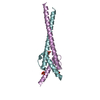
 Movie
Movie Controller


+ Open data

- Basic information
| Entry | Database: PDB / ID: 6g6j | ||||||
|---|---|---|---|---|---|---|---|
| Title | The crystal structures of Human MYC:MAX bHLHZip complex | ||||||
 Components Components |
| ||||||
 Keywords Keywords | APOPTOSIS / Myc/Max | ||||||
| Function / homology |  Function and homology information Function and homology informationMad-Max complex / positive regulation of metanephric cap mesenchymal cell proliferation / positive regulation of acinar cell proliferation / acinar cell proliferation / SCF ubiquitin ligase complex binding / NK T cell proliferation / Myc-Max complex / regulation of somatic stem cell population maintenance / regulation of cell cycle process / RNA polymerase II transcription repressor complex ...Mad-Max complex / positive regulation of metanephric cap mesenchymal cell proliferation / positive regulation of acinar cell proliferation / acinar cell proliferation / SCF ubiquitin ligase complex binding / NK T cell proliferation / Myc-Max complex / regulation of somatic stem cell population maintenance / regulation of cell cycle process / RNA polymerase II transcription repressor complex / Binding of TCF/LEF:CTNNB1 to target gene promoters / cellular response to interferon-alpha / positive regulation of B cell apoptotic process / RUNX3 regulates WNT signaling / TFAP2 (AP-2) family regulates transcription of cell cycle factors / myotube differentiation / negative regulation of transcription initiation by RNA polymerase II / negative regulation of cell division / Regulation of CDH1 mRNA translation by microRNAs / negative regulation of monocyte differentiation / detection of mechanical stimulus involved in sensory perception of sound / response to growth factor / B cell apoptotic process / response to alkaloid / transcription regulator activator activity / Transcription of E2F targets under negative control by DREAM complex / fibroblast apoptotic process / negative regulation of stress-activated MAPK cascade / Regulation of NFE2L2 gene expression / protein-DNA complex disassembly / positive regulation of mesenchymal cell proliferation / skeletal system morphogenesis / regulation of telomere maintenance / branching involved in ureteric bud morphogenesis / Signaling by ALK / middle ear morphogenesis / rRNA metabolic process / negative regulation of gene expression via chromosomal CpG island methylation / E-box binding / pigmentation / Transcriptional Regulation by E2F6 / positive regulation of telomere maintenance / MLL1 complex / skeletal muscle cell differentiation / chromosome organization / positive regulation of transcription initiation by RNA polymerase II / positive regulation of intrinsic apoptotic signaling pathway by p53 class mediator / Cyclin E associated events during G1/S transition / core promoter sequence-specific DNA binding / Cyclin A:Cdk2-associated events at S phase entry / negative regulation of fibroblast proliferation / ERK1 and ERK2 cascade / positive regulation of epithelial cell proliferation / transcription coregulator binding / euchromatin / G1/S transition of mitotic cell cycle / SMAD2/SMAD3:SMAD4 heterotrimer regulates transcription / protein processing / protein-DNA complex / positive regulation of miRNA transcription / DNA-binding transcription repressor activity, RNA polymerase II-specific / MAPK6/MAPK4 signaling / NOTCH1 Intracellular Domain Regulates Transcription / cellular response to xenobiotic stimulus / RNA polymerase II transcription regulator complex / spindle / positive regulation of fibroblast proliferation / intrinsic apoptotic signaling pathway in response to DNA damage / Constitutive Signaling by NOTCH1 PEST Domain Mutants / Constitutive Signaling by NOTCH1 HD+PEST Domain Mutants / Transcriptional regulation of granulopoiesis / Wnt signaling pathway / sequence-specific double-stranded DNA binding / cellular response to UV / MAPK cascade / regulation of gene expression / Interleukin-4 and Interleukin-13 signaling / DNA-binding transcription activator activity, RNA polymerase II-specific / transcription by RNA polymerase II / cellular response to hypoxia / Estrogen-dependent gene expression / DNA-binding transcription factor binding / intracellular iron ion homeostasis / DNA-binding transcription factor activity, RNA polymerase II-specific / protein dimerization activity / Ub-specific processing proteases / nuclear body / RNA polymerase II cis-regulatory region sequence-specific DNA binding / chromatin remodeling / DNA-binding transcription factor activity / response to xenobiotic stimulus / axon / positive regulation of cell population proliferation / DNA damage response / ubiquitin protein ligase binding / positive regulation of gene expression / regulation of transcription by RNA polymerase II / dendrite / negative regulation of apoptotic process / positive regulation of DNA-templated transcription Similarity search - Function | ||||||
| Biological species |  Homo sapiens (human) Homo sapiens (human) | ||||||
| Method |  X-RAY DIFFRACTION / SYNCHROTRON / MOLECULAR REPLACEMENT / Resolution: 2.25 Å X-RAY DIFFRACTION / SYNCHROTRON / MOLECULAR REPLACEMENT / Resolution: 2.25 Å | ||||||
 Authors Authors | Allen, M.D. / Zinzalla, G. | ||||||
 Citation Citation | Journal: Biochemistry / Year: 2019 Title: Crystal Structures and Nuclear Magnetic Resonance Studies of the Apo Form of the c-MYC:MAX bHLHZip Complex Reveal a Helical Basic Region in the Absence of DNA. Authors: Sammak, S. / Hamdani, N. / Gorrec, F. / Allen, M.D. / Freund, S.M.V. / Bycroft, M. / Zinzalla, G. | ||||||
| History |
|
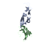
- Structure visualization
| Structure viewer | Molecule: MolmilJmol/JSmol |
|---|
- Downloads & links
-Download
| PDBx/mmCIF format | 6g6j.cif.gz | 81.2 KB | Display | PDBx/mmCIF format |
|---|---|---|---|---|
| PDB format | pdb6g6j.ent.gz | 61.4 KB | Display | PDB format |
| PDBx/mmJSON format | 6g6j.json.gz | Tree view | PDBx/mmJSON format | |
| Others |  Other downloads Other downloads |
-Validation report
| Arichive directory | https://data.pdbj.org/pub/pdb/validation_reports/g6/6g6jftp://data.pdbj.org/pub/pdb/validation_reports/g6/6g6j | HTTPS FTP |
|---|
-Related structure data
| Related structure data |  6g6kC  6g6lC  1knpS S: Starting model for refinement C: citing same article ( |
|---|---|
| Similar structure data |


-Links
 PDBj
PDBj


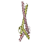
- Assembly
| Deposited unit | 
| ||||||||
|---|---|---|---|---|---|---|---|---|---|
| 1 | 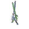
| ||||||||
| 2 | 
| ||||||||
| Unit cell |
|
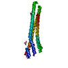
-Components
| #1: Protein | Mass: 11501.192 Da / Num. of mol.: 2 Source method: isolated from a genetically manipulated source Source: (gene. exp.) Homo sapiens (human) / Gene: MYC, BHLHE39 / Production host:  #2: Protein | Mass: 9909.151 Da / Num. of mol.: 2 Source method: isolated from a genetically manipulated source Source: (gene. exp.) Homo sapiens (human) / Gene: MAX, BHLHD4 / Production host: #3: Chemical | ChemComp-SO4 /   Mass: 96.063 Da / Num. of mol.: 6 / Source method: obtained synthetically / Formula: SO4 Mass: 96.063 Da / Num. of mol.: 6 / Source method: obtained synthetically / Formula: SO4#4: Water | ChemComp-HOH / |  Mass: 18.015 Da / Num. of mol.: 180 / Source method: isolated from a natural source / Formula: H2O Mass: 18.015 Da / Num. of mol.: 180 / Source method: isolated from a natural source / Formula: H2O |
|---|
-Experimental details
-Experiment
| Experiment | Method: X-RAY DIFFRACTION / Number of used crystals: 1 |
|---|
- Sample preparation
| Crystal | Density Matthews: 3.06 Å3/Da / Density % sol: 59.74 % |
|---|---|
| Crystal grow | Temperature: 277 K / Method: vapor diffusion, sitting drop / pH: 7 Details: 20% (w/v) PEG 3350 0.2M sodium sulfate decahydrate, pH 7 |
-Data collection
| Diffraction | Mean temperature: 100 K |
|---|---|
| Diffraction source | Source: SYNCHROTRON / Site: Diamond  / Beamline: I03 / Wavelength: 0.97625 Å / Beamline: I03 / Wavelength: 0.97625 Å |
| Detector | Type: DECTRIS PILATUS3 6M / Detector: PIXEL / Date: Aug 8, 2017 |
| Radiation | Protocol: SINGLE WAVELENGTH / Monochromatic (M) / Laue (L): M / Scattering type: x-ray |
| Radiation wavelength | Wavelength: 0.97625 Å / Relative weight: 1 |
| Reflection | Resolution: 2.25→51.93 Å / Num. obs: 25627 / % possible obs: 99.7 % / Redundancy: 4.8 % / CC1/2: 0.997 / Rmerge(I) obs: 0.098 / Rpim(I) all: 0.048 / Rrim(I) all: 0.109 / Net I/σ(I): 8.9 |
| Reflection shell | Resolution: 2.25→2.37 Å / Redundancy: 4.9 % / Rmerge(I) obs: 0.545 / Mean I/σ(I) obs: 2.8 / Num. unique obs: 3679 / CC1/2: 0.875 / Rpim(I) all: 0.265 / Rrim(I) all: 0.608 / % possible all: 99.7 |
- Processing
| Software |
| ||||||||||||||||||||||||||||||||||||||||||||||||||||||||||||||||||||||
|---|---|---|---|---|---|---|---|---|---|---|---|---|---|---|---|---|---|---|---|---|---|---|---|---|---|---|---|---|---|---|---|---|---|---|---|---|---|---|---|---|---|---|---|---|---|---|---|---|---|---|---|---|---|---|---|---|---|---|---|---|---|---|---|---|---|---|---|---|---|---|---|
| Refinement | Method to determine structure: MOLECULAR REPLACEMENT Starting model: 1KNP Resolution: 2.25→40.554 Å / SU ML: 0.23 / Cross valid method: FREE R-VALUE / σ(F): 1.33 / Phase error: 29.53
| ||||||||||||||||||||||||||||||||||||||||||||||||||||||||||||||||||||||
| Solvent computation | Shrinkage radii: 0.9 Å / VDW probe radii: 1.11 Å | ||||||||||||||||||||||||||||||||||||||||||||||||||||||||||||||||||||||
| Refinement step | Cycle: LAST / Resolution: 2.25→40.554 Å
| ||||||||||||||||||||||||||||||||||||||||||||||||||||||||||||||||||||||
| Refine LS restraints |
| ||||||||||||||||||||||||||||||||||||||||||||||||||||||||||||||||||||||
| LS refinement shell |
|
