 Movie
Movie Controller

[English] 日本語
 Yorodumi
- PDB-1ktz: Crystal Structure of the Human TGF-beta Type II Receptor Extracel... -
+ Open data

- Basic information
| Entry | Database: PDB / ID: 1ktz | ||||||
|---|---|---|---|---|---|---|---|
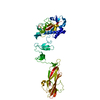
| Title | Crystal Structure of the Human TGF-beta Type II Receptor Extracellular Domain in Complex with TGF-beta3 | ||||||
 Components Components |
| ||||||
 Keywords Keywords | CYTOKINE/CYTOKINE RECEPTOR / CYTOKINE-RECEPTOR COMPLEX / CYTOKINE-CYTOKINE RECEPTOR COMPLEX | ||||||
| Function / homology |  Function and homology information Function and homology informationuterine wall breakdown / positive regulation of tolerance induction to self antigen / positive regulation of B cell tolerance induction / inferior endocardial cushion morphogenesis / transforming growth factor beta receptor activity, type II / bronchus morphogenesis / mammary gland morphogenesis / lens fiber cell apoptotic process / detection of hypoxia / growth plate cartilage chondrocyte growth ...uterine wall breakdown / positive regulation of tolerance induction to self antigen / positive regulation of B cell tolerance induction / inferior endocardial cushion morphogenesis / transforming growth factor beta receptor activity, type II / bronchus morphogenesis / mammary gland morphogenesis / lens fiber cell apoptotic process / detection of hypoxia / growth plate cartilage chondrocyte growth / tricuspid valve morphogenesis / TGFBR2 MSI Frameshift Mutants in Cancer / miRNA transport / transforming growth factor beta ligand-receptor complex / type III transforming growth factor beta receptor binding / aorta morphogenesis / positive regulation of epithelial to mesenchymal transition involved in endocardial cushion formation / Langerhans cell differentiation / transforming growth factor beta receptor activity / TGFBR2 Kinase Domain Mutants in Cancer / cardiac left ventricle morphogenesis / negative regulation of macrophage cytokine production / secondary palate development / SMAD2/3 Phosphorylation Motif Mutants in Cancer / TGFBR1 KD Mutants in Cancer / endocardial cushion fusion / positive regulation of tight junction disassembly / positive regulation of T cell tolerance induction / positive regulation of NK T cell differentiation / membranous septum morphogenesis / TGFBR3 regulates TGF-beta signaling / activin receptor activity, type I / activin receptor complex / regulation of stem cell proliferation / lung lobe morphogenesis / type II transforming growth factor beta receptor binding / receptor protein serine/threonine kinase / activin binding / transmembrane receptor protein serine/threonine kinase activity / TGFBR1 LBD Mutants in Cancer / SMAD protein signal transduction / embryonic cranial skeleton morphogenesis / myeloid dendritic cell differentiation / glycosaminoglycan binding / activin receptor signaling pathway / type I transforming growth factor beta receptor binding / positive regulation of CD4-positive, alpha-beta T cell proliferation / mammary gland development / outflow tract septum morphogenesis / regulation of stem cell differentiation / response to cholesterol / cell-cell junction organization / transforming growth factor beta binding / lens development in camera-type eye / atrioventricular valve morphogenesis / aortic valve morphogenesis / kinase activator activity / face morphogenesis / embryonic hemopoiesis / odontogenesis / artery morphogenesis / positive regulation of filopodium assembly / positive regulation of mesenchymal cell proliferation / Molecules associated with elastic fibres / trachea formation / lung alveolus development / smoothened signaling pathway / branching involved in blood vessel morphogenesis / ventricular septum morphogenesis / blood vessel development / heart looping / SMAD binding / roof of mouth development / outflow tract morphogenesis / TGF-beta receptor signaling activates SMADs / positive regulation of collagen biosynthetic process / positive regulation of cell division / positive regulation of SMAD protein signal transduction / positive regulation of epithelial cell migration / negative regulation of vascular associated smooth muscle cell proliferation / epithelial to mesenchymal transition / ECM proteoglycans / vasculogenesis / positive regulation of epithelial to mesenchymal transition / salivary gland morphogenesis / Notch signaling pathway / positive regulation of stress fiber assembly / response to progesterone / gastrulation / transforming growth factor beta receptor signaling pathway / platelet alpha granule lumen / cytokine activity / Downregulation of TGF-beta receptor signaling / positive regulation of protein secretion / growth factor activity / TGF-beta receptor signaling in EMT (epithelial to mesenchymal transition) / brain development / caveola / cellular response to growth factor stimulus / positive regulation of reactive oxygen species metabolic process Similarity search - Function | ||||||
| Biological species |  Homo sapiens (human) Homo sapiens (human) | ||||||
| Method |  X-RAY DIFFRACTION / SYNCHROTRON / MIR / Resolution: 2.15 Å X-RAY DIFFRACTION / SYNCHROTRON / MIR / Resolution: 2.15 Å | ||||||
 Authors Authors | Hart, P.J. / Deep, S. / Taylor, A.B. / Shu, Z. / Hinck, C.S. / Hinck, A.P. | ||||||
 Citation Citation | Journal: Nat.Struct.Biol. / Year: 2002 Title: Crystal structure of the human TbetaR2 ectodomain--TGF-beta3 complex. Authors: Hart, P.J. / Deep, S. / Taylor, A.B. / Shu, Z. / Hinck, C.S. / Hinck, A.P. | ||||||
| History |
|
- Structure visualization
| Structure viewer | Molecule: MolmilJmol/JSmol |
|---|
- Downloads & links
-Download
| PDBx/mmCIF format | 1ktz.cif.gz | 54.5 KB | Display | PDBx/mmCIF format |
|---|---|---|---|---|
| PDB format | pdb1ktz.ent.gz | 38.8 KB | Display | PDB format |
| PDBx/mmJSON format | 1ktz.json.gz | Tree view | PDBx/mmJSON format | |
| Others |  Other downloads Other downloads |
-Validation report
| Arichive directory | https://data.pdbj.org/pub/pdb/validation_reports/kt/1ktzftp://data.pdbj.org/pub/pdb/validation_reports/kt/1ktz | HTTPS FTP |
|---|
-Related structure data
| Similar structure data |
|---|


-Links
 PDBj
PDBj


- Assembly
| Deposited unit | 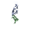
| ||||||||
|---|---|---|---|---|---|---|---|---|---|
| 1 | 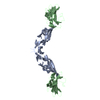
| ||||||||
| Unit cell |
| ||||||||
| Details | The second part of the biological assembly is generated by the crystallographic symmetry operator y, x, -z and a translation of one unit cell length along the z-axis. |
-Components
| #1: Protein | Mass: 12734.504 Da / Num. of mol.: 1 Source method: isolated from a genetically manipulated source Source: (gene. exp.) Homo sapiens (human) / Production host:  |
|---|---|
| #2: Protein | Mass: 13853.632 Da / Num. of mol.: 1 / Fragment: Extracellular Domain Source method: isolated from a genetically manipulated source Source: (gene. exp.) Homo sapiens (human) / Production host: |
| #3: Water | ChemComp-HOH /  Mass: 18.015 Da / Num. of mol.: 163 / Source method: isolated from a natural source / Formula: H2O Mass: 18.015 Da / Num. of mol.: 163 / Source method: isolated from a natural source / Formula: H2O |
| Has protein modification | Y |
-Experimental details
-Experiment
| Experiment | Method: X-RAY DIFFRACTION / Number of used crystals: 1 |
|---|
- Sample preparation
| Crystal | Density Matthews: 5 Å3/Da / Density % sol: 75 % | ||||||||||||||||||||||||
|---|---|---|---|---|---|---|---|---|---|---|---|---|---|---|---|---|---|---|---|---|---|---|---|---|---|
| Crystal grow | Details: HANGING DROP VAPOR DIFFUSION USING A WELL SOLUTION CONTAINING 20% (V/V) 2-METHYL-2,4-PENTANEDIOL AND 0.1 M CITRATE PH 4.0 | ||||||||||||||||||||||||
| Crystal | *PLUS Density % sol: 75 % | ||||||||||||||||||||||||
| Crystal grow | *PLUS pH: 4 / Method: vapor diffusion, hanging drop | ||||||||||||||||||||||||
| Components of the solutions | *PLUS
|
-Data collection
| Diffraction | Mean temperature: 100 K |
|---|---|
| Diffraction source | Source: SYNCHROTRON / Site: NSLS  / Beamline: X8C / Wavelength: 1.1 Å / Beamline: X8C / Wavelength: 1.1 Å |
| Detector | Type: ADSC QUANTUM 4 / Detector: CCD / Date: Nov 7, 2000 |
| Radiation | Protocol: SINGLE WAVELENGTH / Monochromatic (M) / Laue (L): M / Scattering type: x-ray |
| Radiation wavelength | Wavelength: 1.1 Å / Relative weight: 1 |
| Reflection | Resolution: 2.15→26 Å / Num. all: 29006 / Num. obs: 29006 / % possible obs: 99.8 % / Observed criterion σ(I): -3 / Redundancy: 5.3 % / Biso Wilson estimate: 29.4 Å2 / Rmerge(I) obs: 0.058 / Net I/σ(I): 24.5 |
| Reflection shell | Resolution: 2.15→2.23 Å / Redundancy: 4.4 % / Rmerge(I) obs: 0.533 / Mean I/σ(I) obs: 2.7 / Num. unique all: 2824 / % possible all: 98.8 |
| Reflection | *PLUS Lowest resolution: 100 Å / Num. measured all: 154181 |
| Reflection shell | *PLUS % possible obs: 98.8 % |
- Processing
| Software |
| ||||||||||||||||||||||||||||||||||||||||
|---|---|---|---|---|---|---|---|---|---|---|---|---|---|---|---|---|---|---|---|---|---|---|---|---|---|---|---|---|---|---|---|---|---|---|---|---|---|---|---|---|---|
| Refinement | Method to determine structure: MIR / Resolution: 2.15→25.61 Å / Rfactor Rfree error: 0.005 / Data cutoff high absF: 3888970.26 / Data cutoff low absF: 0 / Isotropic thermal model: RESTRAINED / Cross valid method: THROUGHOUT / σ(F): 0 / Stereochemistry target values: Engh & Huber
| ||||||||||||||||||||||||||||||||||||||||
| Solvent computation | Solvent model: FLAT MODEL / Bsol: 61.0368 Å2 / ksol: 0.356719 e/Å3 | ||||||||||||||||||||||||||||||||||||||||
| Displacement parameters | Biso mean: 45.7 Å2
| ||||||||||||||||||||||||||||||||||||||||
| Refine analyze |
| ||||||||||||||||||||||||||||||||||||||||
| Refinement step | Cycle: LAST / Resolution: 2.15→25.61 Å
| ||||||||||||||||||||||||||||||||||||||||
| Refine LS restraints |
| ||||||||||||||||||||||||||||||||||||||||
| LS refinement shell | Resolution: 2.15→2.23 Å / Rfactor Rfree error: 0.02 / Total num. of bins used: 10
| ||||||||||||||||||||||||||||||||||||||||
| Xplor file |
| ||||||||||||||||||||||||||||||||||||||||
| Software | *PLUS Name: CNS / Version: 1 / Classification: refinement | ||||||||||||||||||||||||||||||||||||||||
| Refinement | *PLUS Lowest resolution: 26 Å / σ(F): 0 / % reflection Rfree: 6.9 % | ||||||||||||||||||||||||||||||||||||||||
| Solvent computation | *PLUS | ||||||||||||||||||||||||||||||||||||||||
| Displacement parameters | *PLUS Biso mean: 45.7 Å2 | ||||||||||||||||||||||||||||||||||||||||
| Refine LS restraints | *PLUS
| ||||||||||||||||||||||||||||||||||||||||
| LS refinement shell | *PLUS Rfactor Rfree: 0.288 / % reflection Rfree: 7.1 % / Rfactor Rwork: 0.265 |
