 Movie
Movie Controller

[English] 日本語
 Yorodumi
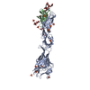
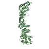
- PDB-3c5x: Crystal structure of the precursor membrane protein- envelope pro... -
+ Open data

- Basic information
| Entry | Database: PDB / ID: 3c5x | |||||||||
|---|---|---|---|---|---|---|---|---|---|---|
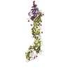
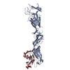
| Title | Crystal structure of the precursor membrane protein- envelope protein heterodimer from the dengue 2 virus at low pH | |||||||||
 Components Components |
| |||||||||
 Keywords Keywords | VIRAL PROTEIN / BETA BARREL / prM-E protein complex / Helicase / Hydrolase / Nucleotide-binding / RNA replication / Transmembrane | |||||||||
| Function / homology |  Function and homology information Function and homology informationhost cell mitochondrion / symbiont-mediated suppression of host JAK-STAT cascade via inhibition of host TYK2 activity / symbiont-mediated suppression of host JAK-STAT cascade via inhibition of STAT2 activity / symbiont-mediated suppression of host cytoplasmic pattern recognition receptor signaling pathway via inhibition of MAVS activity / viral capsid / ribonucleoside triphosphate phosphatase activity / double-stranded RNA binding / channel activity / monoatomic ion transmembrane transport / clathrin-dependent endocytosis of virus by host cell ...host cell mitochondrion / symbiont-mediated suppression of host JAK-STAT cascade via inhibition of host TYK2 activity / symbiont-mediated suppression of host JAK-STAT cascade via inhibition of STAT2 activity / symbiont-mediated suppression of host cytoplasmic pattern recognition receptor signaling pathway via inhibition of MAVS activity / viral capsid / ribonucleoside triphosphate phosphatase activity / double-stranded RNA binding / channel activity / monoatomic ion transmembrane transport / clathrin-dependent endocytosis of virus by host cell / molecular adaptor activity / methyltransferase cap1 activity / mRNA 5'-cap (guanine-N7-)-methyltransferase activity / RNA helicase activity / protein dimerization activity / symbiont-mediated suppression of host innate immune response / host cell perinuclear region of cytoplasm / host cell endoplasmic reticulum membrane / symbiont-mediated suppression of host type I interferon-mediated signaling pathway / serine-type endopeptidase activity / symbiont-mediated activation of host autophagy / viral RNA genome replication / RNA-directed RNA polymerase activity / fusion of virus membrane with host endosome membrane / viral envelope / lipid binding / virion attachment to host cell / host cell nucleus / virion membrane / structural molecule activity / DNA-templated transcription / proteolysis / extracellular region / ATP binding / metal ion binding Similarity search - Function | |||||||||
| Biological species |  Dengue virus 2 Thailand/16681/84Dengue virus 2 Dengue virus 2 Thailand/16681/84Dengue virus 2 | |||||||||
| Method |  X-RAY DIFFRACTION / SYNCHROTRON / MOLECULAR REPLACEMENT / Resolution: 2.2 Å X-RAY DIFFRACTION / SYNCHROTRON / MOLECULAR REPLACEMENT / Resolution: 2.2 Å | |||||||||
 Authors Authors | Li, L. | |||||||||
 Citation Citation | Journal: Science / Year: 2008 Title: The flavivirus precursor membrane-envelope protein complex: structure and maturation. Authors: Long Li / Shee-Mei Lok / I-Mei Yu / Ying Zhang / Richard J Kuhn / Jue Chen / Michael G Rossmann /  Abstract: Many viruses go through a maturation step in the final stages of assembly before being transmitted to another host. The maturation process of flaviviruses is directed by the proteolytic cleavage of ...Many viruses go through a maturation step in the final stages of assembly before being transmitted to another host. The maturation process of flaviviruses is directed by the proteolytic cleavage of the precursor membrane protein (prM), turning inert virus into infectious particles. We have determined the 2.2 angstrom resolution crystal structure of a recombinant protein in which the dengue virus prM is linked to the envelope glycoprotein E. The structure represents the prM-E heterodimer and fits well into the cryo-electron microscopy density of immature virus at neutral pH. The pr peptide beta-barrel structure covers the fusion loop in E, preventing fusion with host cell membranes. The structure provides a basis for identifying the stages of its pH-directed conformational metamorphosis during maturation, ending with release of pr when budding from the host. | |||||||||
| History |
|
- Structure visualization
| Structure viewer | Molecule: MolmilJmol/JSmol |
|---|
- Downloads & links
-Download
| PDBx/mmCIF format | 3c5x.cif.gz | 115.1 KB | Display | PDBx/mmCIF format |
|---|---|---|---|---|
| PDB format | pdb3c5x.ent.gz | 85.3 KB | Display | PDB format |
| PDBx/mmJSON format | 3c5x.json.gz | Tree view | PDBx/mmJSON format | |
| Others |  Other downloads Other downloads |
-Validation report
| Arichive directory | https://data.pdbj.org/pub/pdb/validation_reports/c5/3c5xftp://data.pdbj.org/pub/pdb/validation_reports/c5/3c5x | HTTPS FTP |
|---|
-Related structure data
| Related structure data | 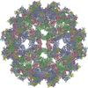 3c6dC  3c6eC 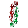 1tg8S C: citing same article ( S: Starting model for refinement |
|---|---|
| Similar structure data |


-Links
 PDBj
PDBj

- Assembly
| Deposited unit | 
| ||||||||
|---|---|---|---|---|---|---|---|---|---|
| 1 |
| ||||||||
| Unit cell |
| ||||||||
| Components on special symmetry positions |
|
-Components
| #1: Protein | Mass: 44801.410 Da / Num. of mol.: 1 / Fragment: UNP residues 281-674 Source method: isolated from a genetically manipulated source Source: (gene. exp.) Dengue virus 2 Thailand/16681/84 / Genus: Flavivirus / Species: Dengue virus / Strain: 16681 / Plasmid: pMT/BiP-prM-TEV-E / Genus (production host): Drosophila / Production host:  |
|---|---|
| #2: Protein | Mass: 14870.891 Da / Num. of mol.: 1 / Fragment: UNP residues 115-244 Source method: isolated from a genetically manipulated source Source: (gene. exp.) Dengue virus 2 / Genus: Flavivirus / Species: Dengue virus / Strain: 2 / Plasmid: pMT/BiP-prM-TEV-E / Genus (production host): Drosophila / Production host: |
| #3: Polysaccharide | 2-acetamido-2-deoxy-alpha-D-glucopyranose-(1-4)-2-acetamido-2-deoxy-beta-D-glucopyranose Source method: isolated from a genetically manipulated source |
| #4: Polysaccharide | beta-D-mannopyranose-(1-3)-[beta-D-mannopyranose-(1-6)]alpha-D-altropyranose-(1-4)-2-acetamido-2- ...beta-D-mannopyranose-(1-3)-[beta-D-mannopyranose-(1-6)]alpha-D-altropyranose-(1-4)-2-acetamido-2-deoxy-beta-D-glucopyranose-(1-4)-2-acetamido-2-deoxy-beta-D-glucopyranose Source method: isolated from a genetically manipulated source |
| #5: Water | ChemComp-HOH /  Mass: 18.015 Da / Num. of mol.: 84 / Source method: isolated from a natural source / Formula: H2O Mass: 18.015 Da / Num. of mol.: 84 / Source method: isolated from a natural source / Formula: H2O |
| Has protein modification | Y |
-Experimental details
-Experiment
| Experiment | Method: X-RAY DIFFRACTION / Number of used crystals: 1 |
|---|
- Sample preparation
| Crystal | Density Matthews: 3.52 Å3/Da / Density % sol: 65.08 % |
|---|---|
| Crystal grow | Temperature: 293 K / Method: vapor diffusion, hanging drop / pH: 5.5 Details: Bis-Tris 0.1M pH5.5, PEG 3350 12%, VAPOR DIFFUSION, HANGING DROP, temperature 293K |
-Data collection
| Diffraction | Mean temperature: 100 K |
|---|---|
| Diffraction source | Source: SYNCHROTRON / Site: APS / Beamline: 23-ID-D / Wavelength: 1.03 Å |
| Detector | Type: MARMOSAIC 300 mm CCD / Detector: CCD / Date: Jun 17, 2007 |
| Radiation | Monochromator: Double crystal cryo-cooled Si(111) / Protocol: SINGLE WAVELENGTH / Monochromatic (M) / Laue (L): M / Scattering type: x-ray |
| Radiation wavelength | Wavelength: 1.03 Å / Relative weight: 1 |
| Reflection | Resolution: 2.2→50 Å / Num. all: 42500 / Num. obs: 42500 / % possible obs: 98.5 % / Redundancy: 6.8 % / Biso Wilson estimate: 48.4 Å2 / Rmerge(I) obs: 0.056 / Rsym value: 0.056 / Net I/σ(I): 30.3 |
| Reflection shell | Resolution: 2.2→2.28 Å / Redundancy: 5.5 % / Rmerge(I) obs: 0.408 / Mean I/σ(I) obs: 3.8 / Num. unique all: 3953 / Rsym value: 0.31 / % possible all: 92.9 |
- Processing
| Software |
| |||||||||||||||||||||||||
|---|---|---|---|---|---|---|---|---|---|---|---|---|---|---|---|---|---|---|---|---|---|---|---|---|---|---|
| Refinement | Method to determine structure: MOLECULAR REPLACEMENT Starting model: PDB ENTRY 1TG8 Resolution: 2.2→50 Å / Isotropic thermal model: anisotropic / Cross valid method: THROUGHOUT / σ(F): 0 / σ(I): 0 / Stereochemistry target values: Engh & Huber
| |||||||||||||||||||||||||
| Displacement parameters |
| |||||||||||||||||||||||||
| Refine analyze |
| |||||||||||||||||||||||||
| Refinement step | Cycle: LAST / Resolution: 2.2→50 Å
| |||||||||||||||||||||||||
| Refine LS restraints |
| |||||||||||||||||||||||||
| LS refinement shell | Resolution: 2.2→2.28 Å
|
