 Movie
Movie Controller


+ Open data

- Basic information
| Entry | Database: PDB / ID: 2y44 | ||||||
|---|---|---|---|---|---|---|---|
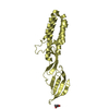
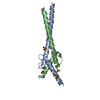
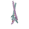
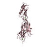
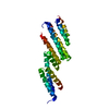
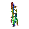
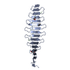
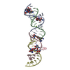
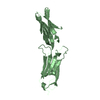
| Title | Crystal structure of GARP from Trypanosoma congolense | ||||||
 Components Components | GLUTAMIC ACID/ALANINE-RICH PROTEIN | ||||||
 Keywords Keywords | MEMBRANE PROTEIN / SURFACE PROTEIN | ||||||
| Function / homology | Trypanosoma glutamic acid/alanine-rich protein / Glutamic acid/alanine-rich protein of Trypanosoma / Ferritin - #80 / Ferritin / Up-down Bundle / Mainly Alpha / IODIDE ION / Alanine-rich surface protein / Glutamic acid/alanine-rich protein Function and homology information Function and homology information | ||||||
| Biological species |  | ||||||
| Method |  X-RAY DIFFRACTION / SAD / Resolution: 1.65 Å X-RAY DIFFRACTION / SAD / Resolution: 1.65 Å | ||||||
 Authors Authors | Loveless, B.C. / Mason, J.W. / Sakurai, T. / Inoue, N. / Razavi, M. / Pearson, T.W. / Boulanger, M.J. | ||||||
 Citation Citation | Journal: J.Biol.Chem. / Year: 2011 Title: Structural Characterization and Epitope Mapping of the Glutamic Acid/Alanine-Rich Protein from Trypanosoma Congolense: Defining Assembly on the Parasite Cell Surface. Authors: Loveless, B.C. / Mason, J.W. / Sakurai, T. / Inoue, N. / Razavi, M. / Pearson, T.W. / Boulanger, M.J. | ||||||
| History |
|
- Structure visualization
| Structure viewer | Molecule: MolmilJmol/JSmol |
|---|
- Downloads & links
-Download
| PDBx/mmCIF format | 2y44.cif.gz | 53 KB | Display | PDBx/mmCIF format |
|---|---|---|---|---|
| PDB format | pdb2y44.ent.gz | 37.4 KB | Display | PDB format |
| PDBx/mmJSON format | 2y44.json.gz | Tree view | PDBx/mmJSON format | |
| Others |  Other downloads Other downloads |
-Validation report
| Arichive directory | https://data.pdbj.org/pub/pdb/validation_reports/y4/2y44ftp://data.pdbj.org/pub/pdb/validation_reports/y4/2y44 | HTTPS FTP |
|---|
-Related structure data
| Similar structure data |
|---|

-Links
 PDBj
PDBj- Assembly
| Deposited unit | 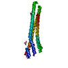
| ||||||||
|---|---|---|---|---|---|---|---|---|---|
| 1 |
| ||||||||
| Unit cell |
|
-Components
| #1: Protein | Mass: 19395.477 Da / Num. of mol.: 1 / Fragment: ECTODOMAIN, RESIDUES 40-224 Source method: isolated from a genetically manipulated source Source: (gene. exp.)  | ||||||||
|---|---|---|---|---|---|---|---|---|---|
| #2: Chemical | ChemComp-IOD /   Mass: 126.904 Da / Num. of mol.: 4 / Source method: obtained synthetically / Formula: I Mass: 126.904 Da / Num. of mol.: 4 / Source method: obtained synthetically / Formula: I#3: Chemical | ChemComp-GOL / |   Mass: 92.094 Da / Num. of mol.: 1 / Source method: obtained synthetically / Formula: C3H8O3 Mass: 92.094 Da / Num. of mol.: 1 / Source method: obtained synthetically / Formula: C3H8O3#4: Water | ChemComp-HOH / |  Mass: 18.015 Da / Num. of mol.: 255 / Source method: isolated from a natural source / Formula: H2O Mass: 18.015 Da / Num. of mol.: 255 / Source method: isolated from a natural source / Formula: H2OHas protein modification | Y | Sequence details | GENE CLONED FROM A SPECIFIC STRAIN AND WITH THE KNOWN LEVEL OF POLYMORPHISM IN THIS FAMILY OF ...GENE CLONED FROM A SPECIFIC STRAIN AND WITH THE KNOWN LEVEL OF POLYMORPHI | |
-Experimental details
-Experiment
| Experiment | Method: X-RAY DIFFRACTION / Number of used crystals: 1 |
|---|
- Sample preparation
| Crystal | Density Matthews: 2.44 Å3/Da / Density % sol: 49.6 % / Description: NONE |
|---|
-Data collection
| Diffraction | Mean temperature: 100 K |
|---|---|
| Diffraction source | Source: ROTATING ANODE / Wavelength: 1.54 |
| Detector | Details: MIRRORS |
| Radiation | Protocol: SINGLE WAVELENGTH / Monochromatic (M) / Laue (L): M / Scattering type: x-ray |
| Radiation wavelength | Wavelength: 1.54 Å / Relative weight: 1 |
| Reflection | Resolution: 1.65→34.62 Å / Num. obs: 20523 / % possible obs: 93.1 % / Observed criterion σ(I): 2 / Redundancy: 7.86 % / Rmerge(I) obs: 0.09 / Net I/σ(I): 10.9 |
| Reflection shell | Resolution: 1.65→1.71 Å / Redundancy: 6.85 % / Rmerge(I) obs: 0.45 / Mean I/σ(I) obs: 3.5 / % possible all: 89 |
- Processing
| Software |
| ||||||||||||||||||||||||||||||||||||||||||||||||||||||||||||||||||||||||||||||||||||||||||||||||||||||||||||||||||||||||||||||||||||||||||||||||||||||||||||||||||||||||||||||||||||||
|---|---|---|---|---|---|---|---|---|---|---|---|---|---|---|---|---|---|---|---|---|---|---|---|---|---|---|---|---|---|---|---|---|---|---|---|---|---|---|---|---|---|---|---|---|---|---|---|---|---|---|---|---|---|---|---|---|---|---|---|---|---|---|---|---|---|---|---|---|---|---|---|---|---|---|---|---|---|---|---|---|---|---|---|---|---|---|---|---|---|---|---|---|---|---|---|---|---|---|---|---|---|---|---|---|---|---|---|---|---|---|---|---|---|---|---|---|---|---|---|---|---|---|---|---|---|---|---|---|---|---|---|---|---|---|---|---|---|---|---|---|---|---|---|---|---|---|---|---|---|---|---|---|---|---|---|---|---|---|---|---|---|---|---|---|---|---|---|---|---|---|---|---|---|---|---|---|---|---|---|---|---|---|---|
| Refinement | Method to determine structure: SAD Starting model: NONE Resolution: 1.65→34.62 Å / Cor.coef. Fo:Fc: 0.951 / Cor.coef. Fo:Fc free: 0.926 / SU B: 2.171 / SU ML: 0.073 / Cross valid method: THROUGHOUT / ESU R: 0.121 / ESU R Free: 0.125 / Stereochemistry target values: MAXIMUM LIKELIHOOD / Details: HYDROGENS HAVE BEEN ADDED IN THE RIDING POSITIONS.
| ||||||||||||||||||||||||||||||||||||||||||||||||||||||||||||||||||||||||||||||||||||||||||||||||||||||||||||||||||||||||||||||||||||||||||||||||||||||||||||||||||||||||||||||||||||||
| Solvent computation | Ion probe radii: 0.8 Å / Shrinkage radii: 0.8 Å / VDW probe radii: 1.4 Å / Solvent model: MASK | ||||||||||||||||||||||||||||||||||||||||||||||||||||||||||||||||||||||||||||||||||||||||||||||||||||||||||||||||||||||||||||||||||||||||||||||||||||||||||||||||||||||||||||||||||||||
| Displacement parameters | Biso mean: 22.995 Å2
| ||||||||||||||||||||||||||||||||||||||||||||||||||||||||||||||||||||||||||||||||||||||||||||||||||||||||||||||||||||||||||||||||||||||||||||||||||||||||||||||||||||||||||||||||||||||
| Refinement step | Cycle: LAST / Resolution: 1.65→34.62 Å
| ||||||||||||||||||||||||||||||||||||||||||||||||||||||||||||||||||||||||||||||||||||||||||||||||||||||||||||||||||||||||||||||||||||||||||||||||||||||||||||||||||||||||||||||||||||||
| Refine LS restraints |
|