 Movie
Movie Controller

[English] 日本語
 Yorodumi
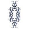
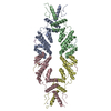
- PDB-5c6q: Crystal structure of the apo TOPLESS related protein 2 (TPR2) N-t... -
+ Open data

- Basic information
| Entry | Database: PDB / ID: 5c6q | ||||||||||||
|---|---|---|---|---|---|---|---|---|---|---|---|---|---|
| Title | Crystal structure of the apo TOPLESS related protein 2 (TPR2) N-terminal domain (1-209) from rice | ||||||||||||
 Components Components | ASPR2 protein | ||||||||||||
 Keywords Keywords | TRANSCRIPTION / Transcriptional corepressor / alpha-helical structure / tetrameric protein / plant transcriptional repression / plant development | ||||||||||||
| Function / homology |  Function and homology information Function and homology informationpositive regulation of pattern recognition receptor signaling pathway / protein sequestering activity / regulation of DNA-templated transcription / plasma membrane Similarity search - Function | ||||||||||||
| Biological species |  | ||||||||||||
| Method |  X-RAY DIFFRACTION / SYNCHROTRON / MOLECULAR REPLACEMENT / Resolution: 3.251 Å X-RAY DIFFRACTION / SYNCHROTRON / MOLECULAR REPLACEMENT / Resolution: 3.251 Å | ||||||||||||
 Authors Authors | Ke, J. / Ma, H. / Gu, X. / Brunzelle, J.S. / Xu, H.E. / Melcher, K. | ||||||||||||
| Funding support |  United States, 3items United States, 3items
| ||||||||||||
 Citation Citation | Journal: Sci Adv / Year: 2015 Title: Structural basis for recognition of diverse transcriptional repressors by the TOPLESS family of corepressors. Authors: Ke, J. / Ma, H. / Gu, X. / Thelen, A. / Brunzelle, J.S. / Li, J. / Xu, H.E. / Melcher, K. | ||||||||||||
| History |
|
- Structure visualization
| Structure viewer | Molecule: MolmilJmol/JSmol |
|---|
- Downloads & links
-Download
| PDBx/mmCIF format | 5c6q.cif.gz | 57.2 KB | Display | PDBx/mmCIF format |
|---|---|---|---|---|
| PDB format | pdb5c6q.ent.gz | 39.5 KB | Display | PDB format |
| PDBx/mmJSON format | 5c6q.json.gz | Tree view | PDBx/mmJSON format | |
| Others |  Other downloads Other downloads |
-Validation report
| Arichive directory | https://data.pdbj.org/pub/pdb/validation_reports/c6/5c6qftp://data.pdbj.org/pub/pdb/validation_reports/c6/5c6q | HTTPS FTP |
|---|
-Related structure data
| Related structure data | 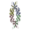 4zheSC 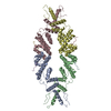 5c6vC 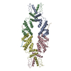 5c7eC  5c7fC S: Starting model for refinement C: citing same article ( |
|---|---|
| Similar structure data |


-Links
 PDBj
PDBj- Assembly
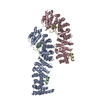
| Deposited unit | 
| ||||||||
|---|---|---|---|---|---|---|---|---|---|
| 1 | 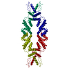
| ||||||||
| Unit cell |
|
-Components
| #1: Protein | Mass: 24903.611 Da / Num. of mol.: 1 / Fragment: N-terminal domain (UNP residues 1-209) Source method: isolated from a genetically manipulated source Source: (gene. exp.)  |
|---|---|
| #2: Chemical | ChemComp-ZN /   Mass: 65.409 Da / Num. of mol.: 1 / Source method: obtained synthetically / Formula: Zn Mass: 65.409 Da / Num. of mol.: 1 / Source method: obtained synthetically / Formula: Zn |
| #3: Water | ChemComp-HOH /  Mass: 18.015 Da / Num. of mol.: 4 / Source method: isolated from a natural source / Formula: H2O Mass: 18.015 Da / Num. of mol.: 4 / Source method: isolated from a natural source / Formula: H2O |
-Experimental details
-Experiment
| Experiment | Method: X-RAY DIFFRACTION / Number of used crystals: 1 |
|---|
- Sample preparation
| Crystal | Density Matthews: 3 Å3/Da / Density % sol: 58.8 % |
|---|---|
| Crystal grow | Temperature: 293 K / Method: vapor diffusion, sitting drop / pH: 6.5 Details: 25% PEG 3350, 0.2 M magnesium chloride, 0.1 M BIS-TRIS, pH 6.5 |
-Data collection
| Diffraction | Mean temperature: 100 K |
|---|---|
| Diffraction source | Source: SYNCHROTRON / Site: APS / Beamline: 21-ID-D / Wavelength: 1.27821 Å |
| Detector | Type: MARMOSAIC 300 mm CCD / Detector: CCD / Date: Oct 30, 2014 |
| Radiation | Monochromator: Ni FILTER / Protocol: SINGLE WAVELENGTH / Monochromatic (M) / Laue (L): M / Scattering type: x-ray |
| Radiation wavelength | Wavelength: 1.27821 Å / Relative weight: 1 |
| Reflection | Resolution: 3.25→50 Å / Num. all: 10544 / Num. obs: 10544 / % possible obs: 100 % / Redundancy: 15.3 % / Net I/σ(I): 27.5 |
| Reflection shell | Resolution: 3.25→3.43 Å / Redundancy: 15.6 % / Mean I/σ(I) obs: 3.5 / % possible all: 100 |
- Processing
| Software |
| ||||||||||||||||||||||||||||
|---|---|---|---|---|---|---|---|---|---|---|---|---|---|---|---|---|---|---|---|---|---|---|---|---|---|---|---|---|---|
| Refinement | Method to determine structure: MOLECULAR REPLACEMENT Starting model: 4ZHE Resolution: 3.251→34.71 Å / SU ML: 0.39 / Cross valid method: FREE R-VALUE / σ(F): 1.57 / Phase error: 32.18 / Stereochemistry target values: ML
| ||||||||||||||||||||||||||||
| Solvent computation | Shrinkage radii: 0.9 Å / VDW probe radii: 1.11 Å / Solvent model: FLAT BULK SOLVENT MODEL | ||||||||||||||||||||||||||||
| Refinement step | Cycle: LAST / Resolution: 3.251→34.71 Å
| ||||||||||||||||||||||||||||
| Refine LS restraints |
| ||||||||||||||||||||||||||||
| LS refinement shell |
|
