 Movie
Movie Controller

[English] 日本語
 Yorodumi
- PDB-6g5r: Structure of the UB2H domain of E.coli PBP1B in complex with LpoB -
+ Open data

- Basic information
| Entry | Database: PDB / ID: 6g5r | ||||||||||||
|---|---|---|---|---|---|---|---|---|---|---|---|---|---|
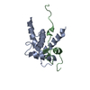
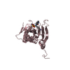
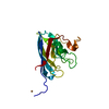
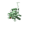
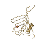
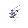
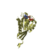
| Title | Structure of the UB2H domain of E.coli PBP1B in complex with LpoB | ||||||||||||
 Components Components | Penicillin-binding protein 1B | ||||||||||||
 Keywords Keywords | TRANSFERASE / Murein polymerase / Complex / LpoB / PBP1B / E. coli / Transpeptidase / Glycosyltransferase / Peptidoglican | ||||||||||||
| Function / homology |  Function and homology information Function and homology informationpositive regulation of bipolar cell growth / cell wall repair / peptidoglycan glycosyltransferase / Action of antimicrobials / peptidoglycan glycosyltransferase activity / serine-type D-Ala-D-Ala carboxypeptidase / serine-type D-Ala-D-Ala carboxypeptidase activity / penicillin binding / peptidoglycan biosynthetic process / peptidoglycan-based cell wall ...positive regulation of bipolar cell growth / cell wall repair / peptidoglycan glycosyltransferase / Action of antimicrobials / peptidoglycan glycosyltransferase activity / serine-type D-Ala-D-Ala carboxypeptidase / serine-type D-Ala-D-Ala carboxypeptidase activity / penicillin binding / peptidoglycan biosynthetic process / peptidoglycan-based cell wall / regulation of cell shape / outer membrane-bounded periplasmic space / response to antibiotic / proteolysis / membrane / plasma membrane Similarity search - Function | ||||||||||||
| Biological species |  | ||||||||||||
| Method | SOLUTION NMR / simulated annealing | ||||||||||||
| Model details | Regulatory domain of Penicillin-Binding Protein | ||||||||||||
 Authors Authors | Simorre, J.P. / Maya Martinez, R.C. / Bougault, C. | ||||||||||||
| Funding support |  France, France,  United Kingdom, 3items United Kingdom, 3items
| ||||||||||||
 Citation Citation | Journal: Mol. Microbiol. / Year: 2018 Title: Induced conformational changes activate the peptidoglycan synthase PBP1B. Authors: Egan, A.J.F. / Maya-Martinez, R. / Ayala, I. / Bougault, C.M. / Banzhaf, M. / Breukink, E. / Vollmer, W. / Simorre, J.P. #1: Journal: J. Biol. Chem. / Year: 2017Title: Structural Insights into Inhibition of Escherichia coli Penicillin-binding Protein 1B. Authors: King, D.T. / Wasney, G.A. / Nosella, M. / Fong, A. / Strynadka, N.C. | ||||||||||||
| History |
|
- Structure visualization
| Structure viewer | Molecule: MolmilJmol/JSmol |
|---|
- Downloads & links
-Download
| PDBx/mmCIF format | 6g5r.cif.gz | 796.9 KB | Display | PDBx/mmCIF format |
|---|---|---|---|---|
| PDB format | pdb6g5r.ent.gz | 680.2 KB | Display | PDB format |
| PDBx/mmJSON format | 6g5r.json.gz | Tree view | PDBx/mmJSON format | |
| Others |  Other downloads Other downloads |
-Validation report
| Arichive directory | https://data.pdbj.org/pub/pdb/validation_reports/g5/6g5rftp://data.pdbj.org/pub/pdb/validation_reports/g5/6g5r | HTTPS FTP |
|---|
-Related structure data
| Related structure data |  6fzkC  6g5sC C: citing same article ( |
|---|---|
| Similar structure data | |
| Other databases |
|


-Links
 PDBj
PDBj
- Assembly
| Deposited unit | 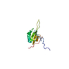
| |||||||||
|---|---|---|---|---|---|---|---|---|---|---|
| 1 |
| |||||||||
| NMR ensembles |
|
 SAXS
SAXS-Components
| #1: Protein | Mass: 13183.969 Da / Num. of mol.: 1 / Fragment: UB2H domain (108-200) Source method: isolated from a genetically manipulated source Details: UB2H is a domain of E. coli of PBP1B (residues 108-200) Source: (gene. exp.) Gene: mrcB, pbpF, ponB, b0149, JW0145 / Plasmid: pET28a / Production host: References: UniProt: P02919, peptidoglycan glycosyltransferase, serine-type D-Ala-D-Ala carboxypeptidase |
|---|
-Experimental details
-Experiment
| Experiment | Method: SOLUTION NMR | ||||||||||||||||||||||||||||||||||||||||||||||||||||||||||||||||||||||||||||||
|---|---|---|---|---|---|---|---|---|---|---|---|---|---|---|---|---|---|---|---|---|---|---|---|---|---|---|---|---|---|---|---|---|---|---|---|---|---|---|---|---|---|---|---|---|---|---|---|---|---|---|---|---|---|---|---|---|---|---|---|---|---|---|---|---|---|---|---|---|---|---|---|---|---|---|---|---|---|---|---|
| NMR experiment |
|
- Sample preparation
| Details |
| ||||||||||||||||||||
|---|---|---|---|---|---|---|---|---|---|---|---|---|---|---|---|---|---|---|---|---|---|
| Sample |
| ||||||||||||||||||||
| Sample conditions | Details: 10 mM Tris/HCl, 200 mM NaCl / Ionic strength: 210 mM / Ionic strength err: 5 / Label: condition_1 / pH: 7.5 / Pressure: 1 atm / Temperature: 293 K |
-NMR measurement
| NMR spectrometer |
|
|---|
- Processing
| NMR software |
| ||||||||||||||||||||||||||||||||
|---|---|---|---|---|---|---|---|---|---|---|---|---|---|---|---|---|---|---|---|---|---|---|---|---|---|---|---|---|---|---|---|---|---|
| Refinement | Method: simulated annealing / Software ordinal: 1 | ||||||||||||||||||||||||||||||||
| NMR representative | Selection criteria: lowest energy | ||||||||||||||||||||||||||||||||
| NMR ensemble | Conformer selection criteria: 20 / Conformers calculated total number: 750 / Conformers submitted total number: 20 |
