 Movie
Movie Controller


+ Open data

- Basic information
| Entry | Database: PDB / ID: 4q6z | ||||||
|---|---|---|---|---|---|---|---|
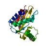
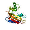
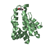
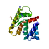
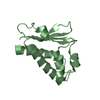
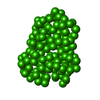
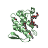
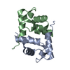
| Title | LpoB C-terminal domain from Escherichia coli | ||||||
 Components Components | Penicillin-binding protein activator LpoB | ||||||
 Keywords Keywords | PROTEIN BINDING / mixed alpha-beta / Lipoprotein / Activator of PBP1b / PBP1b / lipidation / outer-membrane | ||||||
| Function / homology |  Function and homology information Function and homology informationperiplasmic side of cell outer membrane / peptidoglycan biosynthetic process / enzyme regulator activity / regulation of cell shape Similarity search - Function | ||||||
| Biological species |  | ||||||
| Method |  X-RAY DIFFRACTION / SYNCHROTRON / MOLECULAR REPLACEMENT / Resolution: 2.8 Å X-RAY DIFFRACTION / SYNCHROTRON / MOLECULAR REPLACEMENT / Resolution: 2.8 Å | ||||||
 Authors Authors | King, D.T. / Strynadka, N.C.J. | ||||||
 Citation Citation | Journal: J.Biol.Chem. / Year: 2014 Title: Structural Insights into the Lipoprotein Outer Membrane Regulator of Penicillin-binding Protein 1B. Authors: King, D.T. / Lameignere, E. / Strynadka, N.C. | ||||||
| History |
|
- Structure visualization
| Structure viewer | Molecule: MolmilJmol/JSmol |
|---|
- Downloads & links
-Download
| PDBx/mmCIF format | 4q6z.cif.gz | 61.1 KB | Display | PDBx/mmCIF format |
|---|---|---|---|---|
| PDB format | pdb4q6z.ent.gz | 45.6 KB | Display | PDB format |
| PDBx/mmJSON format | 4q6z.json.gz | Tree view | PDBx/mmJSON format | |
| Others |  Other downloads Other downloads |
-Validation report
| Arichive directory | https://data.pdbj.org/pub/pdb/validation_reports/q6/4q6zftp://data.pdbj.org/pub/pdb/validation_reports/q6/4q6z | HTTPS FTP |
|---|
-Related structure data


-Links
 PDBj
PDBj


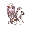
- Assembly
| Deposited unit | 
| ||||||||||||||||||
|---|---|---|---|---|---|---|---|---|---|---|---|---|---|---|---|---|---|---|---|
| 1 | 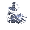
| ||||||||||||||||||
| 2 | 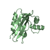
| ||||||||||||||||||
| 3 |
| ||||||||||||||||||
| Unit cell |
| ||||||||||||||||||
| Components on special symmetry positions |
| ||||||||||||||||||
| Noncrystallographic symmetry (NCS) | NCS domain:
NCS domain segments: Component-ID: _ / Ens-ID: 1 / Beg auth comp-ID: TYR / Beg label comp-ID: TYR / End auth comp-ID: GLN / End label comp-ID: GLN / Refine code: _ / Auth seq-ID: 78 - 212 / Label seq-ID: 1 - 135
|
-Components
| #1: Protein | Mass: 14235.036 Da / Num. of mol.: 2 / Fragment: LpoB C-terminal domain Source method: isolated from a genetically manipulated source Source: (gene. exp.) #2: Water | ChemComp-HOH / |  Mass: 18.015 Da / Num. of mol.: 24 / Source method: isolated from a natural source / Formula: H2O Mass: 18.015 Da / Num. of mol.: 24 / Source method: isolated from a natural source / Formula: H2O |
|---|
-Experimental details
-Experiment
| Experiment | Method: X-RAY DIFFRACTION / Number of used crystals: 1 |
|---|
- Sample preparation
| Crystal | Density Matthews: 2.67 Å3/Da / Density % sol: 53.86 % |
|---|---|
| Crystal grow | Temperature: 298.15 K / Method: vapor diffusion, sitting drop / pH: 5.5 Details: 0.1M Bis-Tris, 2M ammonium sulfate , pH 5.5, VAPOR DIFFUSION, SITTING DROP, temperature 298.15K |
-Data collection
| Diffraction | Mean temperature: 100 K |
|---|---|
| Diffraction source | Source: SYNCHROTRON / Site: CLSI  / Beamline: 08B1-1 / Wavelength: 1 Å / Beamline: 08B1-1 / Wavelength: 1 Å |
| Detector | Type: RAYONIX MX300HE / Detector: CCD / Date: Jun 1, 2012 |
| Radiation | Monochromator: KOHZU double crystal monochromator (DCM) / Protocol: SINGLE WAVELENGTH / Monochromatic (M) / Laue (L): M / Scattering type: x-ray |
| Radiation wavelength | Wavelength: 1 Å / Relative weight: 1 |
| Reflection | Resolution: 2.8→68.54 Å / Num. all: 7758 / Num. obs: 7727 / % possible obs: 99.6 % |
| Reflection shell | Resolution: 2.8→2.95 Å / % possible all: 99.3 |
- Processing
| Software |
| |||||||||||||||||||||||||||||||||||||||||||||||||||||||||||||||||||||||||||||||||||||
|---|---|---|---|---|---|---|---|---|---|---|---|---|---|---|---|---|---|---|---|---|---|---|---|---|---|---|---|---|---|---|---|---|---|---|---|---|---|---|---|---|---|---|---|---|---|---|---|---|---|---|---|---|---|---|---|---|---|---|---|---|---|---|---|---|---|---|---|---|---|---|---|---|---|---|---|---|---|---|---|---|---|---|---|---|---|---|
| Refinement | Method to determine structure: MOLECULAR REPLACEMENT / Resolution: 2.8→68.54 Å / Cor.coef. Fo:Fc: 0.915 / Cor.coef. Fo:Fc free: 0.906 / SU B: 21.966 / SU ML: 0.409 / Cross valid method: THROUGHOUT / ESU R Free: 0.428 / Stereochemistry target values: MAXIMUM LIKELIHOOD / Details: HYDROGENS HAVE BEEN ADDED IN THE RIDING POSITIONS
| |||||||||||||||||||||||||||||||||||||||||||||||||||||||||||||||||||||||||||||||||||||
| Solvent computation | Ion probe radii: 0.8 Å / Shrinkage radii: 0.8 Å / VDW probe radii: 1.2 Å / Solvent model: MASK | |||||||||||||||||||||||||||||||||||||||||||||||||||||||||||||||||||||||||||||||||||||
| Displacement parameters | Biso mean: 61.961 Å2
| |||||||||||||||||||||||||||||||||||||||||||||||||||||||||||||||||||||||||||||||||||||
| Refinement step | Cycle: LAST / Resolution: 2.8→68.54 Å
| |||||||||||||||||||||||||||||||||||||||||||||||||||||||||||||||||||||||||||||||||||||
| Refine LS restraints |
| |||||||||||||||||||||||||||||||||||||||||||||||||||||||||||||||||||||||||||||||||||||
| Refine LS restraints NCS | Ens-ID: 1 / Number: 6604 / Refine-ID: X-RAY DIFFRACTION / Type: interatomic distance / Rms dev position: 0.22 Å / Weight position: 0.05
| |||||||||||||||||||||||||||||||||||||||||||||||||||||||||||||||||||||||||||||||||||||
| LS refinement shell | Resolution: 2.8→2.873 Å / Total num. of bins used: 20
|
