 Movie
Movie Controller


+ Open data

- Basic information
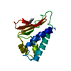
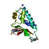
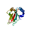
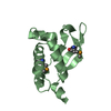
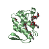
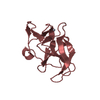
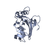
| Entry | Database: PDB / ID: 2z15 | ||||||
|---|---|---|---|---|---|---|---|
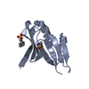
| Title | Crystal structure of human Tob1 protein | ||||||
 Components Components | Protein Tob1 | ||||||
 Keywords Keywords | SIGNALING PROTEIN / human Tob1 protein / Phosphorylation / Structural Genomics / NPPSFA / National Project on Protein Structural and Functional Analyses / RIKEN Structural Genomics/Proteomics Initiative / RSGI | ||||||
| Function / homology |  Function and homology information Function and homology informationregulation of SMAD protein signal transduction / negative regulation of nuclear-transcribed mRNA poly(A) tail shortening / CCR4-NOT complex / positive regulation of nuclear-transcribed mRNA catabolic process, deadenylation-dependent decay / SMAD binding / positive regulation of nuclear-transcribed mRNA poly(A) tail shortening / negative regulation of BMP signaling pathway / negative regulation of osteoblast differentiation / receptor tyrosine kinase binding / transcription corepressor activity ...regulation of SMAD protein signal transduction / negative regulation of nuclear-transcribed mRNA poly(A) tail shortening / CCR4-NOT complex / positive regulation of nuclear-transcribed mRNA catabolic process, deadenylation-dependent decay / SMAD binding / positive regulation of nuclear-transcribed mRNA poly(A) tail shortening / negative regulation of BMP signaling pathway / negative regulation of osteoblast differentiation / receptor tyrosine kinase binding / transcription corepressor activity / regulation of gene expression / negative regulation of translation / negative regulation of cell population proliferation / nucleus / cytoplasm Similarity search - Function | ||||||
| Biological species |  Homo sapiens (human) Homo sapiens (human) | ||||||
| Method |  X-RAY DIFFRACTION / SYNCHROTRON / MAD / Resolution: 2.3 Å X-RAY DIFFRACTION / SYNCHROTRON / MAD / Resolution: 2.3 Å | ||||||
 Authors Authors | Saito, K. / Kishishita, S. / Nishino, A. / Murayama, K. / Terada, T. / Shirouzu, M. / Kigawa, T. / Yokoyama, S. / RIKEN Structural Genomics/Proteomics Initiative (RSGI) | ||||||
 Citation Citation | Journal: To be Published Title: Crystal structure of human Tob1 protein Authors: Saito, K. / Kishishita, S. / Nishino, A. / Murayama, K. / Terada, T. / Shirouzu, M. / Kigawa, T. / Yokoyama, S. | ||||||
| History |
|
- Structure visualization
| Structure viewer | Molecule: MolmilJmol/JSmol |
|---|
- Downloads & links
-Download
| PDBx/mmCIF format | 2z15.cif.gz | 114.8 KB | Display | PDBx/mmCIF format |
|---|---|---|---|---|
| PDB format | pdb2z15.ent.gz | 90.1 KB | Display | PDB format |
| PDBx/mmJSON format | 2z15.json.gz | Tree view | PDBx/mmJSON format | |
| Others |  Other downloads Other downloads |
-Validation report
| Arichive directory | https://data.pdbj.org/pub/pdb/validation_reports/z1/2z15ftp://data.pdbj.org/pub/pdb/validation_reports/z1/2z15 | HTTPS FTP |
|---|
-Related structure data
| Similar structure data | |
|---|---|
| Other databases |

-Links
 PDBj
PDBj- Assembly
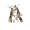
| Deposited unit | 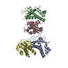
| ||||||||
|---|---|---|---|---|---|---|---|---|---|
| 1 | 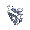
| ||||||||
| 2 | 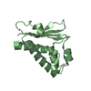
| ||||||||
| 3 | 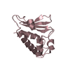
| ||||||||
| 4 | 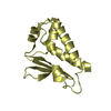
| ||||||||
| Unit cell |
|
-Components
| #1: Protein | Mass: 14636.484 Da / Num. of mol.: 4 / Fragment: UNP residues 1-117 Source method: isolated from a genetically manipulated source Source: (gene. exp.) Homo sapiens (human) / Description: Cell-free protein synthesis / Gene: TOB1, TOB, TROB1 / Plasmid: PK051011-08 / References: UniProt: P50616#2: Water | ChemComp-HOH / |  Mass: 18.015 Da / Num. of mol.: 456 / Source method: isolated from a natural source / Formula: H2O Mass: 18.015 Da / Num. of mol.: 456 / Source method: isolated from a natural source / Formula: H2O |
|---|
-Experimental details
-Experiment
| Experiment | Method: X-RAY DIFFRACTION / Number of used crystals: 1 |
|---|
- Sample preparation
| Crystal | Density Matthews: 2.85 Å3/Da / Density % sol: 56.79 % |
|---|---|
| Crystal grow | Temperature: 293 K / Method: vapor diffusion, hanging drop / pH: 4.8 Details: 0.1M Sodium acetate trihydrate, 8% PEG4000, pH4.8, VAPOR DIFFUSION, HANGING DROP, temperature 293K |
-Data collection
| Diffraction | Mean temperature: 100 K | ||||||||||||
|---|---|---|---|---|---|---|---|---|---|---|---|---|---|
| Diffraction source | Source: SYNCHROTRON / Site: SPring-8  / Beamline: BL26B2 / Wavelength: 0.9789, 0.9794, 0.964 / Beamline: BL26B2 / Wavelength: 0.9789, 0.9794, 0.964 | ||||||||||||
| Detector | Type: RIGAKU JUPITER 210 / Detector: CCD / Date: Oct 6, 2006 / Details: mirrors | ||||||||||||
| Radiation | Monochromator: Si II / Protocol: MAD / Monochromatic (M) / Laue (L): M / Scattering type: x-ray | ||||||||||||
| Radiation wavelength |
| ||||||||||||
| Reflection | Resolution: 2.3→50 Å / Num. obs: 30540 / % possible obs: 99.9 % / Observed criterion σ(I): -3 / Redundancy: 6.76 % / Biso Wilson estimate: 13.7 Å2 / Rsym value: 0.08 / Net I/σ(I): 21.6 | ||||||||||||
| Reflection shell | Resolution: 2.3→2.42 Å / Mean I/σ(I) obs: 4.94 / Rsym value: 0.25 / % possible all: 99.9 |
- Processing
| Software |
| ||||||||||||||||||||||||||||||||||||||||||||||||||||||||||||
|---|---|---|---|---|---|---|---|---|---|---|---|---|---|---|---|---|---|---|---|---|---|---|---|---|---|---|---|---|---|---|---|---|---|---|---|---|---|---|---|---|---|---|---|---|---|---|---|---|---|---|---|---|---|---|---|---|---|---|---|---|---|
| Refinement | Method to determine structure: MAD / Resolution: 2.3→47.44 Å / Rfactor Rfree error: 0.005 / Data cutoff high absF: 70689.48 / Data cutoff low absF: 0 / Isotropic thermal model: RESTRAINED / Cross valid method: THROUGHOUT / σ(F): 0
| ||||||||||||||||||||||||||||||||||||||||||||||||||||||||||||
| Solvent computation | Solvent model: FLAT MODEL / Bsol: 43.413 Å2 / ksol: 0.359178 e/Å3 | ||||||||||||||||||||||||||||||||||||||||||||||||||||||||||||
| Displacement parameters | Biso mean: 25.5 Å2
| ||||||||||||||||||||||||||||||||||||||||||||||||||||||||||||
| Refine analyze |
| ||||||||||||||||||||||||||||||||||||||||||||||||||||||||||||
| Refinement step | Cycle: LAST / Resolution: 2.3→47.44 Å
| ||||||||||||||||||||||||||||||||||||||||||||||||||||||||||||
| Refine LS restraints |
| ||||||||||||||||||||||||||||||||||||||||||||||||||||||||||||
| LS refinement shell | Resolution: 2.3→2.44 Å / Rfactor Rfree error: 0.014 / Total num. of bins used: 6
| ||||||||||||||||||||||||||||||||||||||||||||||||||||||||||||
| Xplor file |
|
