 Movie
Movie Controller

[English] 日本語
 Yorodumi
- PDB-5ubd: Crystal structure of the N-terminal domain (domain 1) of RctB, Rc... -
+ Open data

- Basic information
| Entry | Database: PDB / ID: 5ubd | ||||||
|---|---|---|---|---|---|---|---|
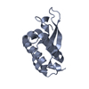
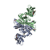
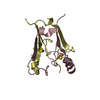
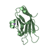
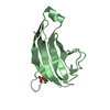
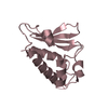
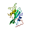
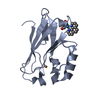
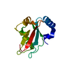
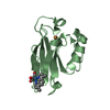
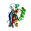
| Title | Crystal structure of the N-terminal domain (domain 1) of RctB, RctB-1-124-L48M | ||||||
 Components Components | RctB replication initiator protein | ||||||
 Keywords Keywords | DNA BINDING PROTEIN / DNA replication initiation / DNA binding / secondary chromosome / Vibrio cholerae | ||||||
| Function / homology | Replication initiator protein RctB, central region / RctB, helix turn helix domain / Vibrionales, replication initiator protein RctB, central region / RctB helix turn helix domain / DUF3346 domain-containing protein / Uncharacterized protein Function and homology information Function and homology information | ||||||
| Biological species |   Vibrio cholerae (bacteria) Vibrio cholerae (bacteria) | ||||||
| Method |  X-RAY DIFFRACTION / SYNCHROTRON / SAD / Resolution: 2.002 Å X-RAY DIFFRACTION / SYNCHROTRON / SAD / Resolution: 2.002 Å | ||||||
 Authors Authors | Orlova, N. / Ivashkiv, O. / Waldor, M.K. / Jeruzalmi, D. | ||||||
| Funding support |  United States, 1items United States, 1items
| ||||||
 Citation Citation | Journal: Nucleic Acids Res. / Year: 2017 Title: The replication initiator of the cholera pathogen's second chromosome shows structural similarity to plasmid initiators. Authors: Orlova, N. / Gerding, M. / Ivashkiv, O. / Olinares, P.D.B. / Chait, B.T. / Waldor, M.K. / Jeruzalmi, D. | ||||||
| History |
|
- Structure visualization
| Structure viewer | Molecule: MolmilJmol/JSmol |
|---|
- Downloads & links
-Download
| PDBx/mmCIF format | 5ubd.cif.gz | 63.1 KB | Display | PDBx/mmCIF format |
|---|---|---|---|---|
| PDB format | pdb5ubd.ent.gz | 45.8 KB | Display | PDB format |
| PDBx/mmJSON format | 5ubd.json.gz | Tree view | PDBx/mmJSON format | |
| Others |  Other downloads Other downloads |
-Validation report
| Arichive directory | https://data.pdbj.org/pub/pdb/validation_reports/ub/5ubdftp://data.pdbj.org/pub/pdb/validation_reports/ub/5ubd | HTTPS FTP |
|---|
-Related structure data

-Links
 PDBj
PDBj- Assembly
| Deposited unit | 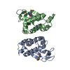
| ||||||||
|---|---|---|---|---|---|---|---|---|---|
| 1 | 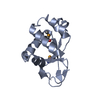
| ||||||||
| 2 | 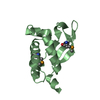
| ||||||||
| Unit cell |
|
-Components
| #1: Protein | Mass: 14998.834 Da / Num. of mol.: 2 / Fragment: domain 1 (UNP residues 1-124) / Mutation: L48M Source method: isolated from a genetically manipulated source Source: (gene. exp.) Vibrio cholerae (bacteria) / Gene: DN30_18, EN12_14105 / Production host: #2: Water | ChemComp-HOH / |  Mass: 18.015 Da / Num. of mol.: 152 / Source method: isolated from a natural source / Formula: H2O Mass: 18.015 Da / Num. of mol.: 152 / Source method: isolated from a natural source / Formula: H2OHas protein modification | Y | |
|---|
-Experimental details
-Experiment
| Experiment | Method: X-RAY DIFFRACTION / Number of used crystals: 1 |
|---|
- Sample preparation
| Crystal | Density Matthews: 2.58 Å3/Da / Density % sol: 52.41 % / Description: plate |
|---|---|
| Crystal grow | Temperature: 277 K / Method: vapor diffusion, sitting drop / pH: 7.5 Details: Crystals of selenomethionine-substituted RctB domain 1 (1 - 124, L48M) were grown by mixing 0.1, or 0.2, or 0.4 uL of the protein solution (20.1 mg/ml RctB-2-124-L48M in 20 mM Tris pH 7.4, ...Details: Crystals of selenomethionine-substituted RctB domain 1 (1 - 124, L48M) were grown by mixing 0.1, or 0.2, or 0.4 uL of the protein solution (20.1 mg/ml RctB-2-124-L48M in 20 mM Tris pH 7.4, 500 mM sodium chloride, 5% glycerol, 5 mM 2-mercaptoethanol) and 0.2 uL of reservoir solution (0.1 M Sodium HEPES pH 7.5, 20% w/v PEG10000). Temp details: cold room |
-Data collection
| Diffraction | Mean temperature: 80 K / Ambient temp details: liquid nitrogen cryo stream |
|---|---|
| Diffraction source | Source: SYNCHROTRON / Site: SSRL / Beamline: BL14-1 / Wavelength: 0.979 Å |
| Detector | Type: MARMOSAIC 325 mm CCD / Detector: CCD / Date: Jun 5, 2015 |
| Radiation | Protocol: SINGLE WAVELENGTH / Monochromatic (M) / Laue (L): M / Scattering type: x-ray |
| Radiation wavelength | Wavelength: 0.979 Å / Relative weight: 1 |
| Reflection | Resolution: 2→50 Å / Num. obs: 19914 / % possible obs: 94.3 % / Redundancy: 2 % / Rsym value: 0.049 / Net I/σ(I): 26.4 |
| Reflection shell | Resolution: 2→2.03 Å / Redundancy: 2 % / Mean I/σ(I) obs: 2.44 / CC1/2: 0.712 / Rsym value: 0.496 / % possible all: 96.9 |
- Processing
| Software |
| ||||||||||||||||||||||||||||||||||||||||||||||||||||||||||||||||||||||||||||||||||||||||||||||||||
|---|---|---|---|---|---|---|---|---|---|---|---|---|---|---|---|---|---|---|---|---|---|---|---|---|---|---|---|---|---|---|---|---|---|---|---|---|---|---|---|---|---|---|---|---|---|---|---|---|---|---|---|---|---|---|---|---|---|---|---|---|---|---|---|---|---|---|---|---|---|---|---|---|---|---|---|---|---|---|---|---|---|---|---|---|---|---|---|---|---|---|---|---|---|---|---|---|---|---|---|
| Refinement | Method to determine structure: SAD / Resolution: 2.002→34.191 Å / SU ML: 0.27 / Cross valid method: FREE R-VALUE / σ(F): 0.18 / Phase error: 31.58
| ||||||||||||||||||||||||||||||||||||||||||||||||||||||||||||||||||||||||||||||||||||||||||||||||||
| Solvent computation | Shrinkage radii: 0.9 Å / VDW probe radii: 1.11 Å | ||||||||||||||||||||||||||||||||||||||||||||||||||||||||||||||||||||||||||||||||||||||||||||||||||
| Refinement step | Cycle: LAST / Resolution: 2.002→34.191 Å
| ||||||||||||||||||||||||||||||||||||||||||||||||||||||||||||||||||||||||||||||||||||||||||||||||||
| Refine LS restraints |
| ||||||||||||||||||||||||||||||||||||||||||||||||||||||||||||||||||||||||||||||||||||||||||||||||||
| LS refinement shell |
|
