[English] 日本語
Yorodumi
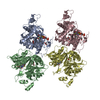
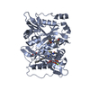
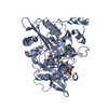
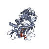
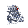
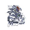
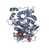
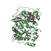
- PDB-6fz5: Human N-myristoyltransferase (NMT1) with Myristoyl-CoA and inhibi... -

+
Open data


ID or keywords:

Loading...

-
Basic information

Entry
Database: PDB / ID: 6fz5
TitleHuman N-myristoyltransferase (NMT1) with Myristoyl-CoA and inhibitor bound
ComponentsGlycylpeptide N-tetradecanoyltransferase 1
KeywordsTRANSFERASE / protein-ligand complex
Function / homology
Function and homology information


myristoyltransferase activity / peptidyl-lysine N6-myristoyltransferase activity / N-terminal peptidyl-glycine N-myristoylation / Late Phase of HIV Life Cycle / cellular ketone metabolic process / regulation of opsin-mediated signaling pathway / Activation, myristolyation of BID and translocation to mitochondria / glycylpeptide N-tetradecanoyltransferase / glycylpeptide N-tetradecanoyltransferase activity / protein localization to membrane ...myristoyltransferase activity / peptidyl-lysine N6-myristoyltransferase activity / N-terminal peptidyl-glycine N-myristoylation / Late Phase of HIV Life Cycle / cellular ketone metabolic process / regulation of opsin-mediated signaling pathway / Activation, myristolyation of BID and translocation to mitochondria / glycylpeptide N-tetradecanoyltransferase / glycylpeptide N-tetradecanoyltransferase activity / protein localization to membrane / Transferases; Acyltransferases; Transferring groups other than aminoacyl groups / Inactivation, recovery and regulation of the phototransduction cascade / in utero embryonic development / plasma membrane / cytosol / cytoplasm
Similarity search - Function
Aminopeptidase - #170 / Glycylpeptide N-tetradecanoyltransferase, conserved site / Myristoyl-CoA:protein N-myristoyltransferase signature 1. / Myristoyl-CoA:protein N-myristoyltransferase signature 2. / Glycylpeptide N-tetradecanoyltransferase / Glycylpeptide N-tetradecanoyltransferase, N-terminal / Glycylpeptide N-tetradecanoyltransferase, C-terminal / Myristoyl-CoA:protein N-myristoyltransferase, N-terminal domain / Myristoyl-CoA:protein N-myristoyltransferase, C-terminal domain / Acyl-CoA N-acyltransferase ...Aminopeptidase - #170 / Glycylpeptide N-tetradecanoyltransferase, conserved site / Myristoyl-CoA:protein N-myristoyltransferase signature 1. / Myristoyl-CoA:protein N-myristoyltransferase signature 2. / Glycylpeptide N-tetradecanoyltransferase / Glycylpeptide N-tetradecanoyltransferase, N-terminal / Glycylpeptide N-tetradecanoyltransferase, C-terminal / Myristoyl-CoA:protein N-myristoyltransferase, N-terminal domain / Myristoyl-CoA:protein N-myristoyltransferase, C-terminal domain / Acyl-CoA N-acyltransferase / Aminopeptidase / 3-Layer(aba) Sandwich / Alpha Beta
Similarity search - Domain/homology
Chem-BXN / TETRADECANOYL-COA / Glycylpeptide N-tetradecanoyltransferase 1
Similarity search - Component
Biological speciesHomo sapiens (human)
MethodX-RAY DIFFRACTION / SYNCHROTRON / MOLECULAR REPLACEMENT / Resolution: 1.89 Å
AuthorsKersten, F.C. / Brenk, R.
Funding support Germany, 1items
OrganizationGrant numberCountry
German Research Foundation252061573 Germany
CitationJournal: J.Med.Chem. / Year: 2020
Title: How To Design Selective Ligands for Highly Conserved Binding Sites: A Case Study UsingN-Myristoyltransferases as a Model System.
Authors: Kersten, C. / Fleischer, E. / Kehrein, J. / Borek, C. / Jaenicke, E. / Sotriffer, C. / Brenk, R.
History
DepositionMar 14, 2018Deposition site: PDBE / Processing site: PDBE
Revision 1.0Mar 27, 2019Provider: repository / Type: Initial release
Revision 1.1Aug 28, 2019Group: Data collection / Database references / Category: citation / citation_author
Item: _citation.country / _citation.journal_abbrev ..._citation.country / _citation.journal_abbrev / _citation.journal_id_ASTM / _citation.journal_id_CSD / _citation.journal_id_ISSN / _citation.pdbx_database_id_DOI / _citation.pdbx_database_id_PubMed / _citation.title / _citation.year
Revision 1.2Mar 18, 2020Group: Database references / Category: citation
Item: _citation.journal_volume / _citation.page_first ..._citation.journal_volume / _citation.page_first / _citation.page_last / _citation.year

-
Structure visualization

Structure viewerMolecule:
MolmilJmol/JSmol

Downloads & links

-
Assembly

Deposited unit
A: Glycylpeptide N-tetradecanoyltransferase 1
B: Glycylpeptide N-tetradecanoyltransferase 1
hetero molecules


Theoretical massNumber of molelcules
Total (without water)92,37012
Polymers88,9692
Non-polymers3,40210
Water6,936385
1
A: Glycylpeptide N-tetradecanoyltransferase 1
hetero molecules


Theoretical massNumber of molelcules
Total (without water)46,1856
Polymers44,4841
Non-polymers1,7015
Water181
TypeNameSymmetry operationNumber
identity operation1_555x,y,z1
2
B: Glycylpeptide N-tetradecanoyltransferase 1
hetero molecules


Theoretical massNumber of molelcules
Total (without water)46,1856
Polymers44,4841
Non-polymers1,7015
Water181
TypeNameSymmetry operationNumber
identity operation1_555x,y,z1
Unit cell
Length a, b, c (Å)92.390, 58.160, 154.001
Angle α, β, γ (deg.)90.00, 92.36, 90.00
Int Tables number5
Space group name H-MC121
Noncrystallographic symmetry (NCS)NCS domain:
IDEns-IDDetails
11A
21B

NCS domain segments:
Dom-IDComponent-IDEns-IDRefine codeAuth asym-IDAuth seq-ID
1010A115 - 496
2010B115 - 496

-
Components

-
Protein , 1 types, 2 molecules AB

#1: Protein Glycylpeptide N-tetradecanoyltransferase 1 / / Myristoyl-CoA:protein N-myristoyltransferase 1 / Type I N-myristoyltransferase / Peptide N- ...Myristoyl-CoA:protein N-myristoyltransferase 1 / Type I N-myristoyltransferase / Peptide N-myristoyltransferase 1


Mass: 44484.277 Da / Num. of mol.: 2
Source method: isolated from a genetically manipulated source
Source: (gene. exp.) Homo sapiens (human) / Gene: NMT1, NMT / Production host: Escherichia coli (E. coli)
References: UniProt: P30419, glycylpeptide N-tetradecanoyltransferase

-
Non-polymers , 5 types, 395 molecules

#2: Chemical ChemComp-BXN / 4-[3-[(8~{a}~{R})-3,4,6,7,8,8~{a}-hexahydro-1~{H}-pyrrolo[1,2-a]pyrazin-2-yl]propyl]-2,6-bis(chloranyl)-~{N}-methyl-~{N}-(1,3,5-trimethylpyrazol-4-yl)benzenesulfonamide


Mass: 514.511 Da / Num. of mol.: 2 / Source method: obtained synthetically / Formula: C23H33Cl2N5O2S / Feature type: SUBJECT OF INVESTIGATION
#3: Chemical ChemComp-MYA / TETRADECANOYL-COA / MYRISTOYL-COA


Mass: 977.890 Da / Num. of mol.: 2 / Source method: obtained synthetically / Formula: C35H62N7O17P3S
#4: Chemical ChemComp-MG / MAGNESIUM ION


Mass: 24.305 Da / Num. of mol.: 2 / Source method: obtained synthetically / Formula: Mg
#5: Chemical
ChemComp-GOL / GLYCEROL / GLYCERIN / PROPANE-1,2,3-TRIOL / Glycerol


Mass: 92.094 Da / Num. of mol.: 4 / Source method: obtained synthetically / Formula: C3H8O3
#6: Water ChemComp-HOH / water / Water


Mass: 18.015 Da / Num. of mol.: 385 / Source method: isolated from a natural source / Formula: H2O

-
Experimental details

-
Experiment

ExperimentMethod: X-RAY DIFFRACTION / Number of used crystals: 1

-
Sample preparation

CrystalDensity Matthews: 2.31 Å3/Da / Density % sol: 46.67 %
Crystal growTemperature: 293 K / Method: vapor diffusion, hanging drop / pH: 4.5
Details: 24% PEG 4000 5 mM NiCl2 0.1 M Na citrate 5% Glycerol

-
Data collection

DiffractionMean temperature: 110 K
Diffraction sourceSource: SYNCHROTRON / Site: ESRF / Beamline: ID29 / Wavelength: 0.976 Å
DetectorType: DECTRIS PILATUS3 6M / Detector: PIXEL / Date: Mar 11, 2018
RadiationProtocol: SINGLE WAVELENGTH / Monochromatic (M) / Laue (L): M / Scattering type: x-ray
Radiation wavelengthWavelength: 0.976 Å / Relative weight: 1
ReflectionResolution: 1.89→76.94 Å / Num. obs: 62185 / % possible obs: 94.8 % / Redundancy: 2.6 % / Biso Wilson estimate: 22.42 Å2 / CC1/2: 0.995 / Rmerge(I) obs: 0.085 / Rpim(I) all: 0.061 / Rrim(I) all: 0.105 / Net I/σ(I): 6.4
Reflection shellResolution: 1.89→1.93 Å / Redundancy: 2.6 % / Rmerge(I) obs: 0.551 / Mean I/σ(I) obs: 1.7 / Num. unique obs: 4069 / CC1/2: 0.605 / Rpim(I) all: 0.403 / Rrim(I) all: 0.687 / % possible all: 96.9

-
Processing

Software
NameVersionClassification
REFMAC5.8.0189refinement
PDB_EXTRACT3.22data extraction
MOSFLM7.2.1data reduction
pointless1.11.4data scaling
Coot0.8.2model building
PHASERphasing
RefinementMethod to determine structure: MOLECULAR REPLACEMENT / Resolution: 1.89→76.94 Å / Cor.coef. Fo:Fc: 0.962 / Cor.coef. Fo:Fc free: 0.946 / SU B: 9.986 / SU ML: 0.14 / Cross valid method: THROUGHOUT / ESU R: 0.17 / ESU R Free: 0.148
Details: HYDROGENS HAVE BEEN ADDED IN THE RIDING POSITIONS #B-factor model selection refi bref ISOT #Solvent related settings scal type SIMP lssc function a sigma n solvent YES solvent vdwprobe 1.0 ...Details: HYDROGENS HAVE BEEN ADDED IN THE RIDING POSITIONS #B-factor model selection refi bref ISOT #Solvent related settings scal type SIMP lssc function a sigma n solvent YES solvent vdwprobe 1.0 ionprobe 0.7 rshrink 0.7 tlsd waters exclude #Restraint weights weight MATRIX 0.03 temp 0.50 #NCS handling ncsr local ncsr align level 0.90 iterate N rmslevel 2.00 ncsr neighbours exclude
RfactorNum. reflection% reflectionSelection details
Rfree0.22339 3212 5.2 %RANDOM
Rwork0.18936 ---
obs0.19115 58973 94.35 %-
Solvent computationIon probe radii: 0.7 Å / Shrinkage radii: 0.7 Å / VDW probe radii: 1 Å
Displacement parametersBiso mean: 33.694 Å2
Baniso -1Baniso -2Baniso -3
1--0.25 Å2-0 Å20.54 Å2
2---0.36 Å2-0 Å2
3---0.57 Å2
Refinement stepCycle: 1 / Resolution: 1.89→76.94 Å
ProteinNucleic acidLigandSolventTotal
Num. atoms6116 0 218 385 6719
Refine LS restraints
Refine-IDTypeDev idealDev ideal targetNumber
X-RAY DIFFRACTIONr_bond_refined_d0.0090.0196525
X-RAY DIFFRACTIONr_bond_other_d0.0020.025960
X-RAY DIFFRACTIONr_angle_refined_deg1.441.9948903
X-RAY DIFFRACTIONr_angle_other_deg0.913313834
X-RAY DIFFRACTIONr_dihedral_angle_1_deg6.2655763
X-RAY DIFFRACTIONr_dihedral_angle_2_deg35.83824.036280
X-RAY DIFFRACTIONr_dihedral_angle_3_deg11.969151035
X-RAY DIFFRACTIONr_dihedral_angle_4_deg13.3351526
X-RAY DIFFRACTIONr_chiral_restr0.0820.2971
X-RAY DIFFRACTIONr_gen_planes_refined0.0060.0217039
X-RAY DIFFRACTIONr_gen_planes_other0.0010.021315
X-RAY DIFFRACTIONr_nbd_refined
X-RAY DIFFRACTIONr_nbd_other
X-RAY DIFFRACTIONr_nbtor_refined
X-RAY DIFFRACTIONr_nbtor_other
X-RAY DIFFRACTIONr_xyhbond_nbd_refined
X-RAY DIFFRACTIONr_xyhbond_nbd_other
X-RAY DIFFRACTIONr_metal_ion_refined
X-RAY DIFFRACTIONr_metal_ion_other
X-RAY DIFFRACTIONr_symmetry_vdw_refined
X-RAY DIFFRACTIONr_symmetry_vdw_other
X-RAY DIFFRACTIONr_symmetry_hbond_refined
X-RAY DIFFRACTIONr_symmetry_hbond_other
X-RAY DIFFRACTIONr_symmetry_metal_ion_refined
X-RAY DIFFRACTIONr_symmetry_metal_ion_other
X-RAY DIFFRACTIONr_mcbond_it1.1421.443047
X-RAY DIFFRACTIONr_mcbond_other1.1421.443048
X-RAY DIFFRACTIONr_mcangle_it1.8472.1513805
X-RAY DIFFRACTIONr_mcangle_other1.8472.1523806
X-RAY DIFFRACTIONr_scbond_it1.4691.6183478
X-RAY DIFFRACTIONr_scbond_other1.4661.6183478
X-RAY DIFFRACTIONr_scangle_it
X-RAY DIFFRACTIONr_scangle_other2.2932.3765097
X-RAY DIFFRACTIONr_long_range_B_refined5.00317.497126
X-RAY DIFFRACTIONr_long_range_B_other4.95917.1027047
X-RAY DIFFRACTIONr_rigid_bond_restr
X-RAY DIFFRACTIONr_sphericity_free
X-RAY DIFFRACTIONr_sphericity_bonded
Refine LS restraints NCS

Ens-ID: 1 / Number: 24970 / Refine-ID: X-RAY DIFFRACTION / Type: interatomic distance / Rms dev position: 0.05 Å / Weight position: 0.05

Dom-IDAuth asym-ID
1A
2B
LS refinement shellResolution: 1.888→1.937 Å / Total num. of bins used: 20
RfactorNum. reflection% reflection
Rfree0.32 209 -
Rwork0.317 4471 -
obs--96.49 %
Refinement TLS params.

Method: refined / Refine-ID: X-RAY DIFFRACTION

IDL112)L122)L132)L222)L232)L332)S11 (Å °)S12 (Å °)S13 (Å °)S21 (Å °)S22 (Å °)S23 (Å °)S31 (Å °)S32 (Å °)S33 (Å °)T112)T122)T132)T222)T232)T332)Origin x (Å)Origin y (Å)Origin z (Å)
10.50410.1371-0.07512.2562-0.16651.00430.051-0.1809-0.02330.2662-0.0092-0.14470.01740.1175-0.04190.2434-0.0088-0.12810.07450.00020.068511.078.84756.287
20.8943-0.10150.06192.2452-0.10251.3258-0.01450.096-0.0696-0.3558-0.04290.2557-0.0775-0.15150.05750.31860.0234-0.14820.0304-0.02340.0798-9.03319.15421.044
Refinement TLS group
IDRefine-IDRefine TLS-IDAuth asym-IDAuth seq-ID
1X-RAY DIFFRACTION1A115 - 1004
2X-RAY DIFFRACTION2B115 - 1004

+
About Yorodumi

-
News

-
Feb 9, 2022. New format data for meta-information of EMDB entries

New format data for meta-information of EMDB entries

  • Version 3 of the EMDB header file is now the official format.
  • The previous official version 1.9 will be removed from the archive.

Related info.:EMDB header

External links:wwPDB to switch to version 3 of the EMDB data model

-
Aug 12, 2020. Covid-19 info

Covid-19 info

URL: https://pdbj.org/emnavi/covid19.php

New page: Covid-19 featured information page in EM Navigator.

Related info.:Covid-19 info / Mar 5, 2020. Novel coronavirus structure data

+
Mar 5, 2020. Novel coronavirus structure data

Novel coronavirus structure data

Related info.:Yorodumi Speices / Aug 12, 2020. Covid-19 info

External links:COVID-19 featured content - PDBj / Molecule of the Month (242):Coronavirus Proteases

+
Jan 31, 2019. EMDB accession codes are about to change! (news from PDBe EMDB page)

EMDB accession codes are about to change! (news from PDBe EMDB page)

  • The allocation of 4 digits for EMDB accession codes will soon come to an end. Whilst these codes will remain in use, new EMDB accession codes will include an additional digit and will expand incrementally as the available range of codes is exhausted. The current 4-digit format prefixed with “EMD-” (i.e. EMD-XXXX) will advance to a 5-digit format (i.e. EMD-XXXXX), and so on. It is currently estimated that the 4-digit codes will be depleted around Spring 2019, at which point the 5-digit format will come into force.
  • The EM Navigator/Yorodumi systems omit the EMD- prefix.

Related info.:Q: What is EMD? / ID/Accession-code notation in Yorodumi/EM Navigator

External links:EMDB Accession Codes are Changing Soon! / Contact to PDBj

+
Jul 12, 2017. Major update of PDB

Major update of PDB

  • wwPDB released updated PDB data conforming to the new PDBx/mmCIF dictionary.
  • This is a major update changing the version number from 4 to 5, and with Remediation, in which all the entries are updated.
  • In this update, many items about electron microscopy experimental information are reorganized (e.g. em_software).
  • Now, EM Navigator and Yorodumi are based on the updated data.

External links:wwPDB Remediation / Enriched Model Files Conforming to OneDep Data Standards Now Available in the PDB FTP Archive

-
Yorodumi

Thousand views of thousand structures

  • Yorodumi is a browser for structure data from EMDB, PDB, SASBDB, etc.
  • This page is also the successor to EM Navigator detail page, and also detail information page/front-end page for Omokage search.
  • The word "yorodu" (or yorozu) is an old Japanese word meaning "ten thousand". "mi" (miru) is to see.

Related info.:EMDB / PDB / SASBDB / Comparison of 3 databanks / Yorodumi Search / Aug 31, 2016. New EM Navigator & Yorodumi / Yorodumi Papers / Jmol/JSmol / Function and homology information / Changes in new EM Navigator and Yorodumi

Read more