 Movie
Movie Controller

[English] 日本語
 Yorodumi
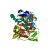
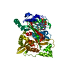
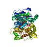
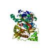
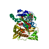
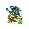
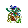
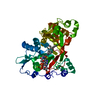
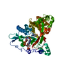
- PDB-6eu5: Leishmania major N-myristoyltransferase with bound myristoyl-CoA ... -
+ Open data

- Basic information
| Entry | Database: PDB / ID: 6eu5 | ||||||
|---|---|---|---|---|---|---|---|
| Title | Leishmania major N-myristoyltransferase with bound myristoyl-CoA and inhibitor | ||||||
 Components Components | Glycylpeptide N-tetradecanoyltransferase | ||||||
 Keywords Keywords | TRANSFERASE / N-Myristoyltransferase / selective inhibitor / parasite | ||||||
| Function / homology |  Function and homology information Function and homology informationglycylpeptide N-tetradecanoyltransferase / glycylpeptide N-tetradecanoyltransferase activity / protein localization to membrane / metal ion binding / cytosol Similarity search - Function | ||||||
| Biological species |  Leishmania major (eukaryote) Leishmania major (eukaryote) | ||||||
| Method |  X-RAY DIFFRACTION / SYNCHROTRON / MOLECULAR REPLACEMENT / Resolution: 1.49608302465 Å X-RAY DIFFRACTION / SYNCHROTRON / MOLECULAR REPLACEMENT / Resolution: 1.49608302465 Å | ||||||
 Authors Authors | Brenk, R. / Kehrein, J. / Kersten, C. | ||||||
| Funding support |  Germany, 1items Germany, 1items
| ||||||
 Citation Citation | Journal: J.Med.Chem. / Year: 2019 Title: How To Design Selective Ligands for Highly Conserved Binding Sites: A Case Study UsingN-Myristoyltransferases as a Model System. Authors: Kersten, C. / Fleischer, E. / Kehrein, J. / Borek, C. / Jaenicke, E. / Sotriffer, C. / Brenk, R. | ||||||
| History |
|
- Structure visualization
| Structure viewer | Molecule: MolmilJmol/JSmol |
|---|
- Downloads & links
-Download
| PDBx/mmCIF format | 6eu5.cif.gz | 217.5 KB | Display | PDBx/mmCIF format |
|---|---|---|---|---|
| PDB format | pdb6eu5.ent.gz | 150 KB | Display | PDB format |
| PDBx/mmJSON format | 6eu5.json.gz | Tree view | PDBx/mmJSON format | |
| Others |  Other downloads Other downloads |
-Validation report
| Arichive directory | https://data.pdbj.org/pub/pdb/validation_reports/eu/6eu5ftp://data.pdbj.org/pub/pdb/validation_reports/eu/6eu5 | HTTPS FTP |
|---|
-Related structure data
| Related structure data |  6ewfC 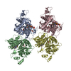 6f56C 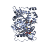 6fz2C 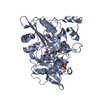 6fz3C 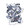 6fz5C  3h5zS S: Starting model for refinement C: citing same article ( |
|---|---|
| Similar structure data |


-Links
 PDBj
PDBj

- Assembly
| Deposited unit | 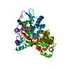
| ||||||||||
|---|---|---|---|---|---|---|---|---|---|---|---|
| 1 |
| ||||||||||
| Unit cell |
|
-Components
| #1: Protein | Mass: 50513.254 Da / Num. of mol.: 1 Source method: isolated from a genetically manipulated source Source: (gene. exp.) Leishmania major (eukaryote) / Gene: NMT, LMJF_32_0080 / Production host:  References: UniProt: Q4Q5S8, glycylpeptide N-tetradecanoyltransferase |
|---|---|
| #2: Chemical | ChemComp-MYA /   Mass: 977.890 Da / Num. of mol.: 1 / Source method: obtained synthetically / Formula: C35H62N7O17P3S Mass: 977.890 Da / Num. of mol.: 1 / Source method: obtained synthetically / Formula: C35H62N7O17P3S |
| #3: Chemical | ChemComp-BXN /   Mass: 514.511 Da / Num. of mol.: 1 / Source method: obtained synthetically / Formula: C23H33Cl2N5O2S / Feature type: SUBJECT OF INVESTIGATION Mass: 514.511 Da / Num. of mol.: 1 / Source method: obtained synthetically / Formula: C23H33Cl2N5O2S / Feature type: SUBJECT OF INVESTIGATION |
| #4: Water | ChemComp-HOH /  Mass: 18.015 Da / Num. of mol.: 227 / Source method: isolated from a natural source / Formula: H2O Mass: 18.015 Da / Num. of mol.: 227 / Source method: isolated from a natural source / Formula: H2O |
-Experimental details
-Experiment
| Experiment | Method: X-RAY DIFFRACTION / Number of used crystals: 1 |
|---|
- Sample preparation
| Crystal | Density Matthews: 2.19 Å3/Da / Density % sol: 43.8 % |
|---|---|
| Crystal grow | Temperature: 293.15 K / Method: vapor diffusion, hanging drop / pH: 5.6 Details: PEG 1500 25 % Sodium chloride 0.2 M Sodium cacodylate 0.1 M |
-Data collection
| Diffraction | Mean temperature: 100 K |
|---|---|
| Diffraction source | Source: SYNCHROTRON / Site: ESRF  / Beamline: MASSIF-1 / Wavelength: 0.966 Å / Beamline: MASSIF-1 / Wavelength: 0.966 Å |
| Detector | Type: DECTRIS PILATUS3 2M / Detector: PIXEL / Date: Sep 29, 2017 |
| Radiation | Monochromator: C(110) / Protocol: SINGLE WAVELENGTH / Monochromatic (M) / Laue (L): M / Scattering type: x-ray |
| Radiation wavelength | Wavelength: 0.966 Å / Relative weight: 1 |
| Reflection | Resolution: 1.496→48.748 Å / Num. obs: 65618 / % possible obs: 96.3 % / Redundancy: 3.1 % / Biso Wilson estimate: 18.630988436 Å2 / CC1/2: 0.999 / Rmerge(I) obs: 0.036 / Rpim(I) all: 0.036 / Rrim(I) all: 0.05 / Net I/σ(I): 16.8 |
| Reflection shell | Resolution: 1.496→1.522 Å / Redundancy: 2.8 % / Rmerge(I) obs: 0.526 / Mean I/σ(I) obs: 2.1 / Num. unique obs: 3245 / CC1/2: 0.751 / Rpim(I) all: 0.526 / Rrim(I) all: 0.744 / % possible all: 95.1 |
- Processing
| Software |
| |||||||||||||||||||||||||||||||||||||||||||||||||||||||||||||||||||||||||||||||||||||||||||||||||||||||||||||||||||||||||||||||||||||||||||||||||||||||||||||||||
|---|---|---|---|---|---|---|---|---|---|---|---|---|---|---|---|---|---|---|---|---|---|---|---|---|---|---|---|---|---|---|---|---|---|---|---|---|---|---|---|---|---|---|---|---|---|---|---|---|---|---|---|---|---|---|---|---|---|---|---|---|---|---|---|---|---|---|---|---|---|---|---|---|---|---|---|---|---|---|---|---|---|---|---|---|---|---|---|---|---|---|---|---|---|---|---|---|---|---|---|---|---|---|---|---|---|---|---|---|---|---|---|---|---|---|---|---|---|---|---|---|---|---|---|---|---|---|---|---|---|---|---|---|---|---|---|---|---|---|---|---|---|---|---|---|---|---|---|---|---|---|---|---|---|---|---|---|---|---|---|---|---|---|
| Refinement | Method to determine structure: MOLECULAR REPLACEMENT Starting model: 3H5Z Resolution: 1.49608302465→48.7479801992 Å / SU ML: 0.165468736412 / Cross valid method: FREE R-VALUE / σ(F): 1.35027070076 / Phase error: 21.0274788376
| |||||||||||||||||||||||||||||||||||||||||||||||||||||||||||||||||||||||||||||||||||||||||||||||||||||||||||||||||||||||||||||||||||||||||||||||||||||||||||||||||
| Solvent computation | Shrinkage radii: 0.9 Å / VDW probe radii: 1.11 Å | |||||||||||||||||||||||||||||||||||||||||||||||||||||||||||||||||||||||||||||||||||||||||||||||||||||||||||||||||||||||||||||||||||||||||||||||||||||||||||||||||
| Displacement parameters | Biso mean: 24.6128418489 Å2 | |||||||||||||||||||||||||||||||||||||||||||||||||||||||||||||||||||||||||||||||||||||||||||||||||||||||||||||||||||||||||||||||||||||||||||||||||||||||||||||||||
| Refinement step | Cycle: LAST / Resolution: 1.49608302465→48.7479801992 Å
| |||||||||||||||||||||||||||||||||||||||||||||||||||||||||||||||||||||||||||||||||||||||||||||||||||||||||||||||||||||||||||||||||||||||||||||||||||||||||||||||||
| Refine LS restraints |
| |||||||||||||||||||||||||||||||||||||||||||||||||||||||||||||||||||||||||||||||||||||||||||||||||||||||||||||||||||||||||||||||||||||||||||||||||||||||||||||||||
| LS refinement shell |
|
