 Movie
Movie Controller

[English] 日本語
 Yorodumi
- PDB-6fvn: DNA polymerase sliding clamp from Mycobacterium tuberculosis with... -
+ Open data

- Basic information
| Entry | Database: PDB / ID: 6fvn | ||||||
|---|---|---|---|---|---|---|---|
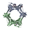
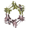
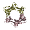
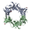
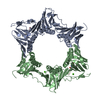
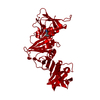
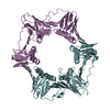
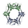
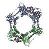
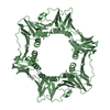
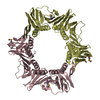
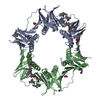
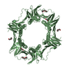
| Title | DNA polymerase sliding clamp from Mycobacterium tuberculosis with bound P7 peptide | ||||||
 Components Components |
| ||||||
 Keywords Keywords | DNA BINDING PROTEIN / DNA sliding clamp | ||||||
| Function / homology |  Function and homology information Function and homology informationDNA polymerase III complex / DNA strand elongation involved in DNA replication / 3'-5' exonuclease activity / DNA-directed DNA polymerase activity / DNA binding / identical protein binding / cytoplasm Similarity search - Function | ||||||
| Biological species |   Mycobacterium tuberculosis (bacteria) Mycobacterium tuberculosis (bacteria)synthetic construct (others) | ||||||
| Method |  X-RAY DIFFRACTION / SYNCHROTRON / MOLECULAR REPLACEMENT / molecular replacement / Resolution: 3.142 Å X-RAY DIFFRACTION / SYNCHROTRON / MOLECULAR REPLACEMENT / molecular replacement / Resolution: 3.142 Å | ||||||
 Authors Authors | Martiel, I. / Andre, C. / Olieric, V. / Guichard, G. / Burnouf, D. | ||||||
| Funding support |  France, 1items France, 1items
| ||||||
 Citation Citation | Journal: Acs Infect Dis. / Year: 2019 Title: Peptide Interactions on Bacterial Sliding Clamps. Authors: Andre, C. / Martiel, I. / Wolff, P. / Landolfo, M. / Lorber, B. / Silva da Veiga, C. / Dejaegere, A. / Dumas, P. / Guichard, G. / Olieric, V. / Wagner, J.G. / Burnouf, D.Y. | ||||||
| History |
|
- Structure visualization
| Structure viewer | Molecule: MolmilJmol/JSmol |
|---|
- Downloads & links
-Download
| PDBx/mmCIF format | 6fvn.cif.gz | 280.7 KB | Display | PDBx/mmCIF format |
|---|---|---|---|---|
| PDB format | pdb6fvn.ent.gz | 222.6 KB | Display | PDB format |
| PDBx/mmJSON format | 6fvn.json.gz | Tree view | PDBx/mmJSON format | |
| Others |  Other downloads Other downloads |
-Validation report
| Arichive directory | https://data.pdbj.org/pub/pdb/validation_reports/fv/6fvnftp://data.pdbj.org/pub/pdb/validation_reports/fv/6fvn | HTTPS FTP |
|---|
-Related structure data
| Related structure data | 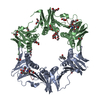 6fvlC 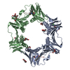 6fvmC  6fvoC 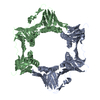 4tr7S S: Starting model for refinement C: citing same article ( |
|---|---|
| Similar structure data |


-Links
 PDBj
PDBj
- Assembly
| Deposited unit | 
| |||||||||||||||||||||||||||||||||||||||||||||||||||||||||||||||||||||||||||||||
|---|---|---|---|---|---|---|---|---|---|---|---|---|---|---|---|---|---|---|---|---|---|---|---|---|---|---|---|---|---|---|---|---|---|---|---|---|---|---|---|---|---|---|---|---|---|---|---|---|---|---|---|---|---|---|---|---|---|---|---|---|---|---|---|---|---|---|---|---|---|---|---|---|---|---|---|---|---|---|---|---|
| 1 | 
| |||||||||||||||||||||||||||||||||||||||||||||||||||||||||||||||||||||||||||||||
| 2 | 
| |||||||||||||||||||||||||||||||||||||||||||||||||||||||||||||||||||||||||||||||
| Unit cell |
| |||||||||||||||||||||||||||||||||||||||||||||||||||||||||||||||||||||||||||||||
| Noncrystallographic symmetry (NCS) | NCS domain:
NCS domain segments:
|