 Movie
Movie Controller

[English] 日本語
 Yorodumi
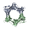
- PDB-5agv: The sliding clamp of Mycobacterium tuberculosis in complex with a... -
+ Open data

- Basic information
| Entry | Database: PDB / ID: 5agv | |||||||||||||||
|---|---|---|---|---|---|---|---|---|---|---|---|---|---|---|---|---|
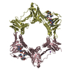
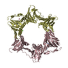
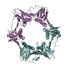
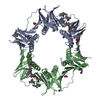
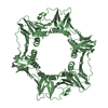
| Title | The sliding clamp of Mycobacterium tuberculosis in complex with a natural product. | |||||||||||||||
 Components Components |
| |||||||||||||||
 Keywords Keywords | TRANSFERASE/ANTIBIOTIC / TRANSFERASE-ANTIBIOTIC COMPLEX / TRANSFERASE / DNAN / NATURAL PRODUCT / SLIDING CLAMP | |||||||||||||||
| Function / homology |  Function and homology information Function and homology informationDNA polymerase III complex / DNA strand elongation involved in DNA replication / 3'-5' exonuclease activity / peptidoglycan-based cell wall / DNA-directed DNA polymerase activity / response to antibiotic / DNA binding / extracellular region / identical protein binding / cytoplasm Similarity search - Function | |||||||||||||||
| Biological species |  MYCOBACTERIUM TUBERCULOSIS H37RV (bacteria)STREPTOMYCES CAELICUS (bacteria) MYCOBACTERIUM TUBERCULOSIS H37RV (bacteria)STREPTOMYCES CAELICUS (bacteria) | |||||||||||||||
| Method |  X-RAY DIFFRACTION / SYNCHROTRON / MOLECULAR REPLACEMENT / Resolution: 1.93 Å X-RAY DIFFRACTION / SYNCHROTRON / MOLECULAR REPLACEMENT / Resolution: 1.93 Å | |||||||||||||||
 Authors Authors | Lukat, P. / Kling, A. / Heinz, D.W. / Mueller, R. | |||||||||||||||
 Citation Citation | Journal: Science / Year: 2015 Title: Antibiotics. Targeting Dnan for Tuberculosis Therapy Using Novel Griselimycins. Authors: Kling, A. / Lukat, P. / Almeida, D.V. / Bauer, A. / Fontaine, E. / Sordello, S. / Zaburannyi, N. / Herrmann, J. / Wenzel, S.C. / Konig, C. / Ammerman, N.C. / Barrio, M.B. / Borchers, K. / ...Authors: Kling, A. / Lukat, P. / Almeida, D.V. / Bauer, A. / Fontaine, E. / Sordello, S. / Zaburannyi, N. / Herrmann, J. / Wenzel, S.C. / Konig, C. / Ammerman, N.C. / Barrio, M.B. / Borchers, K. / Bordon-Pallier, F. / Bronstrup, M. / Courtemanche, G. / Gerlitz, M. / Geslin, M. / Hammann, P. / Heinz, D.W. / Hoffmann, H. / Klieber, S. / Kohlmann, M. / Kurz, M. / Lair, C. / Matter, H. / Nuermberger, E. / Tyagi, S. / Fraisse, L. / Grosset, J.H. / Lagrange, S. / Muller, R. | |||||||||||||||
| History |
|
- Structure visualization
| Structure viewer | Molecule: MolmilJmol/JSmol |
|---|
- Downloads & links
-Download
| PDBx/mmCIF format | 5agv.cif.gz | 348.8 KB | Display | PDBx/mmCIF format |
|---|---|---|---|---|
| PDB format | pdb5agv.ent.gz | 280.5 KB | Display | PDB format |
| PDBx/mmJSON format | 5agv.json.gz | Tree view | PDBx/mmJSON format | |
| Others |  Other downloads Other downloads |
-Validation report
| Arichive directory | https://data.pdbj.org/pub/pdb/validation_reports/ag/5agvftp://data.pdbj.org/pub/pdb/validation_reports/ag/5agv | HTTPS FTP |
|---|
-Related structure data
| Related structure data | 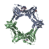 5aguC  5ah2C 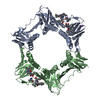 5ah4C 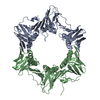 3p16S C: citing same article ( S: Starting model for refinement |
|---|---|
| Similar structure data |


-Links
 PDBj
PDBj
- Assembly
| Deposited unit | 
| ||||||||
|---|---|---|---|---|---|---|---|---|---|
| 1 | 
| ||||||||
| 2 | 
| ||||||||
| Unit cell |
| ||||||||
| Noncrystallographic symmetry (NCS) | NCS oper: (Code: given Matrix: (-0.99984, 0.01309, 0.01194), Vector: |
-Components
-Protein / Protein/peptide , 2 types, 4 molecules ABCD
| #1: Protein | Mass: 42479.207 Da / Num. of mol.: 2 Source method: isolated from a genetically manipulated source Source: (gene. exp.) MYCOBACTERIUM TUBERCULOSIS H37RV (bacteria)Plasmid: PVP008_DNANMTB / Production host: References: UniProt: I6XU56, UniProt: P9WNU1*PLUS, DNA-directed DNA polymerase #2: Protein/peptide | Type: Peptide-like / Class: Antibiotic / Mass: 1197.593 Da / Num. of mol.: 2 / Source method: isolated from a natural source / Source: (natural) STREPTOMYCES CAELICUS (bacteria) / References: BIRD: PRD_002466 |
|---|
-Non-polymers , 4 types, 998 molecules 


| #3: Chemical | ChemComp-CA /  Mass: 40.078 Da / Num. of mol.: 6 / Source method: obtained synthetically / Formula: Ca Mass: 40.078 Da / Num. of mol.: 6 / Source method: obtained synthetically / Formula: Ca#4: Chemical | ChemComp-NA /  Mass: 22.990 Da / Num. of mol.: 12 / Source method: obtained synthetically / Formula: Na Mass: 22.990 Da / Num. of mol.: 12 / Source method: obtained synthetically / Formula: Na#5: Chemical | ChemComp-BU3 / (  Mass: 90.121 Da / Num. of mol.: 5 / Source method: obtained synthetically / Formula: C4H10O2 Mass: 90.121 Da / Num. of mol.: 5 / Source method: obtained synthetically / Formula: C4H10O2#6: Water | ChemComp-HOH / | Mass: 18.015 Da / Num. of mol.: 975 / Source method: isolated from a natural source / Formula: H2O |
|---|
-Details
| Compound details | Novel synthetic analog of natural product Griselimycin |
|---|---|
| Nonpolymer details | CYCLOHEXYLGRISELIMYCIN (CGM): CYCLOHEXYLGRISELIMYCIN IS A SYNTHETICALLY GENERATED DERIVATIVE OF ...CYCLOHEXYL |
| Sequence details | FIRST 4 N-TERMINAL RESIDUES ARE REMAINING FRIOM THE PROTEASE CLEAVAGE. |
-Experimental details
-Experiment
| Experiment | Method: X-RAY DIFFRACTION / Number of used crystals: 1 |
|---|
- Sample preparation
| Crystal | Density Matthews: 2.92 Å3/Da / Density % sol: 57.9 % / Description: NONE |
|---|---|
| Crystal grow | pH: 7.5 Details: 333 MM CACL2, 144 MM LITHIUM ACETATE, 11.7 % (W/V) PEG 8000, 100 MM HEPES-NAOH PH 7.58 |
-Data collection
| Diffraction | Mean temperature: 100 K |
|---|---|
| Diffraction source | Source: SYNCHROTRON / Site: BESSY  / Beamline: 14.1 / Wavelength: 0.91844 / Beamline: 14.1 / Wavelength: 0.91844 |
| Detector | Type: DECTRIS PILATUS 6M / Detector: PIXEL / Date: Sep 24, 2014 / Details: MIRRORS |
| Radiation | Monochromator: KMC-1 / Protocol: SINGLE WAVELENGTH / Monochromatic (M) / Laue (L): M / Scattering type: x-ray |
| Radiation wavelength | Wavelength: 0.91844 Å / Relative weight: 1 |
| Reflection | Resolution: 1.93→40.8 Å / Num. obs: 79001 / % possible obs: 100 % / Observed criterion σ(I): -3 / Redundancy: 6.8 % / Biso Wilson estimate: 25.93 Å2 / Rmerge(I) obs: 0.1 / Net I/σ(I): 13.7 |
| Reflection shell | Resolution: 1.93→1.94 Å / Redundancy: 6.6 % / Rmerge(I) obs: 0.82 / Mean I/σ(I) obs: 2.2 / % possible all: 99.9 |
- Processing
| Software |
| ||||||||||||||||||||||||||||||||||||||||||||||||||||||||||||||||||||||||||||||||||||||||||||||||||||||||||||||||||||||||||||||||||||||||||||||||||||||||||||||||||||||||||||||||||||||||||||||||||||
|---|---|---|---|---|---|---|---|---|---|---|---|---|---|---|---|---|---|---|---|---|---|---|---|---|---|---|---|---|---|---|---|---|---|---|---|---|---|---|---|---|---|---|---|---|---|---|---|---|---|---|---|---|---|---|---|---|---|---|---|---|---|---|---|---|---|---|---|---|---|---|---|---|---|---|---|---|---|---|---|---|---|---|---|---|---|---|---|---|---|---|---|---|---|---|---|---|---|---|---|---|---|---|---|---|---|---|---|---|---|---|---|---|---|---|---|---|---|---|---|---|---|---|---|---|---|---|---|---|---|---|---|---|---|---|---|---|---|---|---|---|---|---|---|---|---|---|---|---|---|---|---|---|---|---|---|---|---|---|---|---|---|---|---|---|---|---|---|---|---|---|---|---|---|---|---|---|---|---|---|---|---|---|---|---|---|---|---|---|---|---|---|---|---|---|---|---|---|
| Refinement | Method to determine structure: MOLECULAR REPLACEMENT Starting model: WWPDB ENTRY 3P16 Resolution: 1.93→40.8 Å / SU ML: 0.2 / σ(F): 1.34 / Phase error: 20.76 / Stereochemistry target values: ML Details: WEAKLY DEFINED SIDECHAINS ARE MODELLED AS ALANINES.
| ||||||||||||||||||||||||||||||||||||||||||||||||||||||||||||||||||||||||||||||||||||||||||||||||||||||||||||||||||||||||||||||||||||||||||||||||||||||||||||||||||||||||||||||||||||||||||||||||||||
| Solvent computation | Shrinkage radii: 0.9 Å / VDW probe radii: 1.11 Å / Solvent model: FLAT BULK SOLVENT MODEL | ||||||||||||||||||||||||||||||||||||||||||||||||||||||||||||||||||||||||||||||||||||||||||||||||||||||||||||||||||||||||||||||||||||||||||||||||||||||||||||||||||||||||||||||||||||||||||||||||||||
| Displacement parameters | Biso mean: 34.47 Å2 | ||||||||||||||||||||||||||||||||||||||||||||||||||||||||||||||||||||||||||||||||||||||||||||||||||||||||||||||||||||||||||||||||||||||||||||||||||||||||||||||||||||||||||||||||||||||||||||||||||||
| Refinement step | Cycle: LAST / Resolution: 1.93→40.8 Å
| ||||||||||||||||||||||||||||||||||||||||||||||||||||||||||||||||||||||||||||||||||||||||||||||||||||||||||||||||||||||||||||||||||||||||||||||||||||||||||||||||||||||||||||||||||||||||||||||||||||
| Refine LS restraints |
| ||||||||||||||||||||||||||||||||||||||||||||||||||||||||||||||||||||||||||||||||||||||||||||||||||||||||||||||||||||||||||||||||||||||||||||||||||||||||||||||||||||||||||||||||||||||||||||||||||||
| LS refinement shell |
| ||||||||||||||||||||||||||||||||||||||||||||||||||||||||||||||||||||||||||||||||||||||||||||||||||||||||||||||||||||||||||||||||||||||||||||||||||||||||||||||||||||||||||||||||||||||||||||||||||||
| Refinement TLS params. | Method: refined / Refine-ID: X-RAY DIFFRACTION
| ||||||||||||||||||||||||||||||||||||||||||||||||||||||||||||||||||||||||||||||||||||||||||||||||||||||||||||||||||||||||||||||||||||||||||||||||||||||||||||||||||||||||||||||||||||||||||||||||||||
| Refinement TLS group |
|
