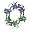
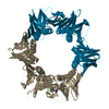
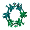
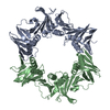
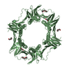
DNA polymerase III complex / DNA strand elongation involved in DNA replication / 3'-5' exonuclease activity / DNA-directed DNA polymerase activity / DNA binding / cytoplasm Similarity search - Function
DNA Polymerase III; Chain A, domain 2 / DNA Polymerase III, subunit A, domain 2 / DNA polymerase III, beta sliding clamp / DNA polymerase III, beta sliding clamp, N-terminal / DNA polymerase III, beta sliding clamp, C-terminal / DNA polymerase III, beta sliding clamp, central / DNA polymerase III beta subunit, N-terminal domain / DNA polymerase III beta subunit, central domain / DNA polymerase III beta subunit, C-terminal domain / DNA polymerase III beta subunit ...DNA Polymerase III; Chain A, domain 2 / DNA Polymerase III, subunit A, domain 2 / DNA polymerase III, beta sliding clamp / DNA polymerase III, beta sliding clamp, N-terminal / DNA polymerase III, beta sliding clamp, C-terminal / DNA polymerase III, beta sliding clamp, central / DNA polymerase III beta subunit, N-terminal domain / DNA polymerase III beta subunit, central domain / DNA polymerase III beta subunit, C-terminal domain / DNA polymerase III beta subunit / : / Roll / Alpha Beta Similarity search - Domain/homology
Resolution: 1.95→50 Å / Cor.coef. Fo:Fc: 0.963 / Cor.coef. Fo:Fc free: 0.945 / SU B: 7.764 / SU ML: 0.099 / Cross valid method: THROUGHOUT / ESU R: 0.213 / ESU R Free: 0.128 / Details: HYDROGENS HAVE BEEN ADDED IN THE RIDING POSITIONS
Rfactor
Num. reflection
% reflection
Selection details
Rfree
0.219
3737
5.04 %
RANDOM
Rwork
0.171
-
-
-
obs
-
74116
99.75 %
-
Solvent computation
Ion probe radii: 0.8 Å / Shrinkage radii: 0.8 Å / VDW probe radii: 1.2 Å
 Movie
Movie Controller

 Yorodumi
 Open data
 Basic information
 Components
 Keywords
 Function and homology information
 Caulobacter vibrioides CB15 (bacteria)
 X-RAY DIFFRACTION /
  Authors
 China, 3items
  Citation
 Structure visualization
 Downloads & links
 Other downloads
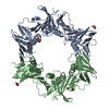


 PDBj
PDBj
 Assembly


 Mass: 92.094 Da / Num. of mol.: 1 / Source method: obtained synthetically / Formula: C3H8O3
 Mass: 62.068 Da / Num. of mol.: 5 / Source method: obtained synthetically / Formula: C2H6O2
 Mass: 282.331 Da / Num. of mol.: 1 / Source method: obtained synthetically / Formula: C12H26O7 / Comment: precipitant*YM
 Mass: 106.120 Da / Num. of mol.: 1 / Source method: obtained synthetically / Formula: C4H10O3
 Mass: 150.173 Da / Num. of mol.: 1 / Source method: obtained synthetically / Formula: C6H14O4
 Sample preparation
 Processing