 Movie
Movie Controller


+ Open data

- Basic information
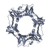
| Entry | Database: PDB / ID: 5g4q | ||||||
|---|---|---|---|---|---|---|---|
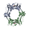
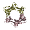
| Title | H.pylori Beta clamp in complex with 5-chloroisatin | ||||||
 Components Components | DNA POLYMERASE III SUBUNIT BETA | ||||||
 Keywords Keywords | TRANSFERASE / DNA SLIDING CLAMP / PROCESSIVITY PROMOTING FACTOR | ||||||
| Function / homology |  Function and homology information Function and homology informationDNA polymerase III complex / DNA strand elongation involved in DNA replication / 3'-5' exonuclease activity / DNA-directed DNA polymerase activity / DNA binding / identical protein binding / cytoplasm Similarity search - Function | ||||||
| Biological species |   HELICOBACTER PYLORI (bacteria) HELICOBACTER PYLORI (bacteria) | ||||||
| Method |  X-RAY DIFFRACTION / MOLECULAR REPLACEMENT / Resolution: 2.3 Å X-RAY DIFFRACTION / MOLECULAR REPLACEMENT / Resolution: 2.3 Å | ||||||
 Authors Authors | Pandey, P. / Gourinath, S. | ||||||
 Citation Citation | Journal: Antibiotics (Basel) / Year: 2018 Title: Screening of E. coli beta-clamp Inhibitors Revealed that Few Inhibit Helicobacter pylori More Effectively: Structural and Functional Characterization. Authors: Pandey, P. / Verma, V. / Dhar, S.K. / Gourinath, S. #1: Journal: Sci.Rep. / Year: 2016Title: Structural Insight Into Beta-Clamp and its Interaction with DNA Ligase in Helicobacter Pylori. Authors: Pandey, P. / Tarique, K.F. / Mazumder, M. / Rehman, S.A.A. / Kumari, N. / Gourinath, S. | ||||||
| History |
|
- Structure visualization
| Structure viewer | Molecule: MolmilJmol/JSmol |
|---|
- Downloads & links
-Download
| PDBx/mmCIF format | 5g4q.cif.gz | 149.7 KB | Display | PDBx/mmCIF format |
|---|---|---|---|---|
| PDB format | pdb5g4q.ent.gz | 118.2 KB | Display | PDB format |
| PDBx/mmJSON format | 5g4q.json.gz | Tree view | PDBx/mmJSON format | |
| Others |  Other downloads Other downloads |
-Validation report
| Arichive directory | https://data.pdbj.org/pub/pdb/validation_reports/g4/5g4qftp://data.pdbj.org/pub/pdb/validation_reports/g4/5g4q | HTTPS FTP |
|---|
-Related structure data
| Related structure data | 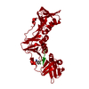 5fveC  5fxtC 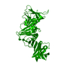 4rkiS S: Starting model for refinement C: citing same article ( |
|---|---|
| Similar structure data |


-Links
 PDBj
PDBj
- Assembly
| Deposited unit | 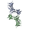
| ||||||||
|---|---|---|---|---|---|---|---|---|---|
| 1 | 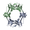
| ||||||||
| 2 | 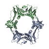
| ||||||||
| Unit cell |
|
-Components
| #1: Protein | Mass: 42238.441 Da / Num. of mol.: 2 Source method: isolated from a genetically manipulated source Details: BETA CLAMP IN COMPLEX WITH 5-CHLOROISATIN / Source: (gene. exp.) HELICOBACTER PYLORI (bacteria) / Strain: 26695 / Organ: STOMACH / Production host: #2: Chemical | ChemComp-2HQ / |   Mass: 181.576 Da / Num. of mol.: 1 / Source method: obtained synthetically / Formula: C8H4ClNO2 Mass: 181.576 Da / Num. of mol.: 1 / Source method: obtained synthetically / Formula: C8H4ClNO2#3: Water | ChemComp-HOH / |  Mass: 18.015 Da / Num. of mol.: 57 / Source method: isolated from a natural source / Formula: H2O Mass: 18.015 Da / Num. of mol.: 57 / Source method: isolated from a natural source / Formula: H2O |
|---|
-Experimental details
-Experiment
| Experiment | Method: X-RAY DIFFRACTION / Number of used crystals: 1 |
|---|
- Sample preparation
| Crystal | Density Matthews: 2.32 Å3/Da / Density % sol: 47 % Description: THIS DATA WAS OBTAINED FROM CRYSTALS SOAKED IN LIGAND BEFORE DIFFRACTION LEADING TO HIGH TEMPERATURE FACTOR AND LOW OCCUPANCY |
|---|---|
| Crystal grow | Details: 6% W/V PEG 20,000, 20% V/V PEG 550MME, 0.01M MGCL2, 0.1M MOPS/ HEPES-NA PH7.3, 0.2M AMMONIUM CITRATE |
-Data collection
| Diffraction | Mean temperature: 287 K |
|---|---|
| Diffraction source | Source: ROTATING ANODE / Wavelength: 0.95 |
| Detector | Type: MARRESEARCH / Detector: CCD / Date: Nov 21, 2015 |
| Radiation | Protocol: SINGLE WAVELENGTH / Monochromatic (M) / Laue (L): M / Scattering type: x-ray |
| Radiation wavelength | Wavelength: 0.95 Å / Relative weight: 1 |
| Reflection | Resolution: 2.3→50 Å / Num. obs: 39777 / % possible obs: 91.1 % / Observed criterion σ(I): 0.3 / Redundancy: 3.6 % / Rmerge(I) obs: 0.05 / Net I/σ(I): 20.2 |
| Reflection shell | Resolution: 2.3→50 Å / Redundancy: 3.4 % / Rmerge(I) obs: 0.35 / Mean I/σ(I) obs: 2.2 / % possible all: 92.8 |
- Processing
| Software | Name: REFMAC / Version: 5 / Classification: refinement | ||||||||||||||||||||
|---|---|---|---|---|---|---|---|---|---|---|---|---|---|---|---|---|---|---|---|---|---|
| Refinement | Method to determine structure: MOLECULAR REPLACEMENT Starting model: PDB NETRY 4RKI Resolution: 2.3→80 Å / Cross valid method: THROUGHOUT / Stereochemistry target values: MAXIMUM LIKELIHOOD Details: HYDROGENS HAVE BEEN ADDED IN THE RIDING POSITIONS. DATA OBTAINED FROM CRYSTALS SOAKED WITH LIGAND LEADING TO HIGH TEMPERATURE FACTOR AND LOW OCCUPANCY.
| ||||||||||||||||||||
| Refinement step | Cycle: LAST / Resolution: 2.3→80 Å
|
