- PDB-5fxt: Crystal Structure of Helicobacter pylori beta clamp in complex wi... -
+
Open data
ID or keywords:
Loading...
-
Basic information
Entry
Database: PDB / ID: 5fxt
Title
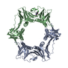
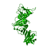
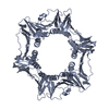
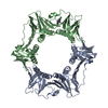
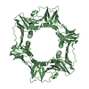
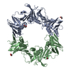
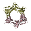
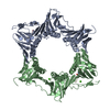
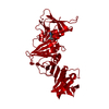
Crystal Structure of Helicobacter pylori beta clamp in complex with Carprofen
Components
DNA POLYMERASE III SUBUNIT BETA
Keywords
TRANSFERASE / DNA CLAMP / SLIDING CLAMP
Function / homology
Function and homology information
DNA polymerase III complex / DNA strand elongation involved in DNA replication / 3'-5' exonuclease activity / DNA-directed DNA polymerase activity / DNA binding / identical protein binding / cytoplasm Similarity search - Function
DNA Polymerase III; Chain A, domain 2 / DNA Polymerase III, subunit A, domain 2 / DNA polymerase III, beta sliding clamp / DNA polymerase III, beta sliding clamp, N-terminal / DNA polymerase III, beta sliding clamp, C-terminal / DNA polymerase III, beta sliding clamp, central / DNA polymerase III beta subunit, N-terminal domain / DNA polymerase III beta subunit, central domain / DNA polymerase III beta subunit, C-terminal domain / DNA polymerase III beta subunit ...DNA Polymerase III; Chain A, domain 2 / DNA Polymerase III, subunit A, domain 2 / DNA polymerase III, beta sliding clamp / DNA polymerase III, beta sliding clamp, N-terminal / DNA polymerase III, beta sliding clamp, C-terminal / DNA polymerase III, beta sliding clamp, central / DNA polymerase III beta subunit, N-terminal domain / DNA polymerase III beta subunit, central domain / DNA polymerase III beta subunit, C-terminal domain / DNA polymerase III beta subunit / : / Roll / Alpha Beta Similarity search - Domain/homology
Resolution: 1.97→74.76 Å / Cor.coef. Fo:Fc: 0.96 / Cor.coef. Fo:Fc free: 0.933 / SU B: 12.067 / SU ML: 0.145 / Cross valid method: THROUGHOUT / ESU R: 0.175 / ESU R Free: 0.169 / Stereochemistry target values: MAXIMUM LIKELIHOOD / Details: HYDROGENS HAVE BEEN ADDED IN THE RIDING POSITIONS.
Rfactor
Num. reflection
% reflection
Selection details
Rfree
0.26049
1557
5.1 %
RANDOM
Rwork
0.20731
-
-
-
obs
0.21009
29254
99.02 %
-
Solvent computation
Ion probe radii: 0.8 Å / Shrinkage radii: 0.8 Å / VDW probe radii: 1.2 Å / Solvent model: MASK
 Movie
Movie Controller

 Yorodumi
 Open data
 Basic information
 Components
 Keywords
 Function and homology information
 HELICOBACTER PYLORI (bacteria)
 X-RAY DIFFRACTION /
  Authors
 Citation
 Structure visualization
 Downloads & links
 Other downloads
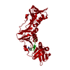


 PDBj
PDBj
 Assembly
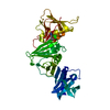

 Mass: 273.714 Da / Num. of mol.: 1 / Source method: obtained synthetically / Formula: C15H12ClNO2
 Mass: 18.015 Da / Num. of mol.: 74 / Source method: isolated from a natural source / Formula: H2O
 Sample preparation
 / Beamline: BM14 / Wavelength: 0.935
  Processing