 Movie
Movie Controller

[English] 日本語
 Yorodumi
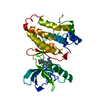
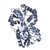
- PDB-6fqp: Crystal structure of TALE homeobox domain transcription factor TG... -
+ Open data

- Basic information
| Entry | Database: PDB / ID: 6fqp | |||||||||
|---|---|---|---|---|---|---|---|---|---|---|
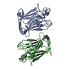
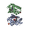
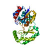
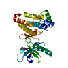
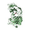
| Title | Crystal structure of TALE homeobox domain transcription factor TGIF1 with its consensus DNA | |||||||||
 Components Components |
| |||||||||
 Keywords Keywords | TRANSCRIPTION / homeobox / three-amino acid loop extension / TGF-beta pathway | |||||||||
| Function / homology |  Function and homology information Function and homology informationpositive regulation of amacrine cell differentiation / regulation of gastrulation / amacrine cell differentiation / nodal signaling pathway / co-SMAD binding / negative regulation of retinoic acid receptor signaling pathway / dorsal/ventral pattern formation / determination of left/right symmetry / neural tube closure / Downregulation of SMAD2/3:SMAD4 transcriptional activity ...positive regulation of amacrine cell differentiation / regulation of gastrulation / amacrine cell differentiation / nodal signaling pathway / co-SMAD binding / negative regulation of retinoic acid receptor signaling pathway / dorsal/ventral pattern formation / determination of left/right symmetry / neural tube closure / Downregulation of SMAD2/3:SMAD4 transcriptional activity / SMAD2/SMAD3:SMAD4 heterotrimer regulates transcription / DNA-binding transcription repressor activity, RNA polymerase II-specific / cellular response to growth factor stimulus / positive regulation of fibroblast proliferation / sequence-specific double-stranded DNA binding / fibroblast proliferation / DNA-binding transcription factor activity, RNA polymerase II-specific / RNA polymerase II cis-regulatory region sequence-specific DNA binding / DNA-binding transcription factor activity / response to xenobiotic stimulus / negative regulation of cell population proliferation / negative regulation of gene expression / chromatin binding / chromatin / negative regulation of transcription by RNA polymerase II / nucleoplasm Similarity search - Function | |||||||||
| Biological species |  Homo sapiens (human) Homo sapiens (human) | |||||||||
| Method |  X-RAY DIFFRACTION / SYNCHROTRON / MOLECULAR REPLACEMENT / Resolution: 2.42 Å X-RAY DIFFRACTION / SYNCHROTRON / MOLECULAR REPLACEMENT / Resolution: 2.42 Å | |||||||||
 Authors Authors | Guca, E. / Macias, M.J. | |||||||||
| Funding support |  Spain, 2items Spain, 2items
| |||||||||
 Citation Citation | Journal: Nucleic Acids Res. / Year: 2018 Title: TGIF1 homeodomain interacts with Smad MH1 domain and represses TGF-beta signaling. Authors: Guca, E. / Sunol, D. / Ruiz, L. / Konkol, A. / Cordero, J. / Torner, C. / Aragon, E. / Martin-Malpartida, P. / Riera, A. / Macias, M.J. | |||||||||
| History |
|
- Structure visualization
| Structure viewer | Molecule: MolmilJmol/JSmol |
|---|
- Downloads & links
-Download
| PDBx/mmCIF format | 6fqp.cif.gz | 94.2 KB | Display | PDBx/mmCIF format |
|---|---|---|---|---|
| PDB format | pdb6fqp.ent.gz | 67.9 KB | Display | PDB format |
| PDBx/mmJSON format | 6fqp.json.gz | Tree view | PDBx/mmJSON format | |
| Others |  Other downloads Other downloads |
-Validation report
| Arichive directory | https://data.pdbj.org/pub/pdb/validation_reports/fq/6fqpftp://data.pdbj.org/pub/pdb/validation_reports/fq/6fqp | HTTPS FTP |
|---|
-Related structure data
| Related structure data |  6fqqC  4xrmS S: Starting model for refinement C: citing same article ( |
|---|---|
| Similar structure data |


-Links
 PDBj
PDBj


- Assembly
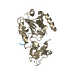
| Deposited unit | 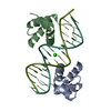
| ||||||||
|---|---|---|---|---|---|---|---|---|---|
| 1 |
| ||||||||
| Unit cell |
|
-Components
| #1: DNA chain | Mass: 4897.204 Da / Num. of mol.: 2 / Source method: obtained synthetically / Source: (synth.) Homo sapiens (human)#2: Protein | Mass: 11729.339 Da / Num. of mol.: 2 Source method: isolated from a genetically manipulated source Details: N-terminal 'GP' sequence comes from the purification tag Source: (gene. exp.) Homo sapiens (human) / Gene: TGIF1, TGIF / Production host:  #3: Chemical | ChemComp-CA / |   Mass: 40.078 Da / Num. of mol.: 1 / Source method: obtained synthetically / Formula: Ca Mass: 40.078 Da / Num. of mol.: 1 / Source method: obtained synthetically / Formula: Ca#4: Water | ChemComp-HOH / |  Mass: 18.015 Da / Num. of mol.: 68 / Source method: isolated from a natural source / Formula: H2O Mass: 18.015 Da / Num. of mol.: 68 / Source method: isolated from a natural source / Formula: H2O |
|---|
-Experimental details
-Experiment
| Experiment | Method: X-RAY DIFFRACTION / Number of used crystals: 1 |
|---|
- Sample preparation
| Crystal | Density Matthews: 3.39 Å3/Da / Density % sol: 63.69 % |
|---|---|
| Crystal grow | Temperature: 277 K / Method: vapor diffusion, sitting drop Details: 0.1M sodium acetate trihydrate pH 4.5, 30% v/v PEG 1500 |
-Data collection
| Diffraction | Mean temperature: 100 K |
|---|---|
| Diffraction source | Source: SYNCHROTRON / Site: ESRF  / Beamline: ID23-1 / Wavelength: 0.972422 Å / Beamline: ID23-1 / Wavelength: 0.972422 Å |
| Detector | Type: DECTRIS PILATUS 6M-F / Detector: PIXEL / Date: May 10, 2017 |
| Radiation | Protocol: SINGLE WAVELENGTH / Monochromatic (M) / Laue (L): M / Scattering type: x-ray |
| Radiation wavelength | Wavelength: 0.972422 Å / Relative weight: 1 |
| Reflection | Resolution: 2.42→67.25 Å / Num. obs: 13814 / % possible obs: 98.9 % / Redundancy: 2 % / Rmerge(I) obs: 0.0275 / Net I/σ(I): 17.3 |
| Reflection shell | Resolution: 2.42→2.51 Å / Rmerge(I) obs: 0.232 / % possible all: 98.7 |
- Processing
| Software |
| ||||||||||||||||||
|---|---|---|---|---|---|---|---|---|---|---|---|---|---|---|---|---|---|---|---|
| Refinement | Method to determine structure: MOLECULAR REPLACEMENT Starting model: 4XRM Resolution: 2.42→45.537 Å / SU ML: 0.28 / Cross valid method: FREE R-VALUE / σ(F): 1.34 / Phase error: 31.24
| ||||||||||||||||||
| Solvent computation | Shrinkage radii: 0.9 Å / VDW probe radii: 1.11 Å | ||||||||||||||||||
| Displacement parameters | Biso max: 117.63 Å2 / Biso mean: 52.9948 Å2 / Biso min: 23.84 Å2 | ||||||||||||||||||
| Refinement step | Cycle: LAST / Resolution: 2.42→45.537 Å
|
