 Movie
Movie Controller


+ Open data

- Basic information
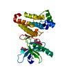
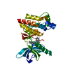
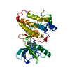
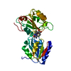
| Entry | Database: PDB / ID: 7kpl | ||||||
|---|---|---|---|---|---|---|---|
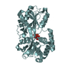
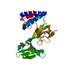
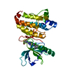
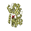
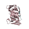
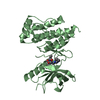
| Title | Crystal structure of hEphB1 in apo form | ||||||
 Components Components | Ephrin type-B receptor 1 | ||||||
 Keywords Keywords | TRANSFERASE / HEPHB1 | ||||||
| Function / homology |  Function and homology information Function and homology informationskeletal muscle satellite cell activation / hindbrain tangential cell migration / negative regulation of skeletal muscle satellite cell proliferation / optic nerve morphogenesis / central nervous system projection neuron axonogenesis / axon guidance receptor activity / transmembrane-ephrin receptor activity / negative regulation of satellite cell differentiation / dendritic spine development / immunological synapse formation ...skeletal muscle satellite cell activation / hindbrain tangential cell migration / negative regulation of skeletal muscle satellite cell proliferation / optic nerve morphogenesis / central nervous system projection neuron axonogenesis / axon guidance receptor activity / transmembrane-ephrin receptor activity / negative regulation of satellite cell differentiation / dendritic spine development / immunological synapse formation / filopodium tip / camera-type eye morphogenesis / dendritic spine morphogenesis / positive regulation of synapse assembly / EPH-Ephrin signaling / neural precursor cell proliferation / retinal ganglion cell axon guidance / Ephrin signaling / regulation of JNK cascade / establishment of cell polarity / cell-substrate adhesion / detection of temperature stimulus involved in sensory perception of pain / EPH-ephrin mediated repulsion of cells / ephrin receptor signaling pathway / regulation of ERK1 and ERK2 cascade / neurogenesis / EPHB-mediated forward signaling / axon guidance / cell chemotaxis / receptor protein-tyrosine kinase / modulation of chemical synaptic transmission / protein autophosphorylation / angiogenesis / early endosome membrane / membrane raft / axon / dendrite / protein-containing complex binding / glutamatergic synapse / endoplasmic reticulum / extracellular exosome / extracellular region / ATP binding / plasma membrane / cytosol Similarity search - Function | ||||||
| Biological species |  Homo sapiens (human) Homo sapiens (human) | ||||||
| Method |  X-RAY DIFFRACTION / SYNCHROTRON / MOLECULAR REPLACEMENT / Resolution: 2.705 Å X-RAY DIFFRACTION / SYNCHROTRON / MOLECULAR REPLACEMENT / Resolution: 2.705 Å | ||||||
 Authors Authors | Ahmed, M. / Wang, P. / Sadek, H. | ||||||
 Citation Citation | Journal: Proc.Natl.Acad.Sci.USA / Year: 2021 Title: Identification of tetracycline combinations as EphB1 tyrosine kinase inhibitors for treatment of neuropathic pain. Authors: Ahmed, M.S. / Wang, P. / Nguyen, N.U.N. / Nakada, Y. / Menendez-Montes, I. / Ismail, M. / Bachoo, R. / Henkemeyer, M. / Sadek, H.A. / Kandil, E.S. | ||||||
| History |
|
- Structure visualization
| Structure viewer | Molecule: MolmilJmol/JSmol |
|---|
- Downloads & links
-Download
| PDBx/mmCIF format | 7kpl.cif.gz | 66.8 KB | Display | PDBx/mmCIF format |
|---|---|---|---|---|
| PDB format | pdb7kpl.ent.gz | 46.9 KB | Display | PDB format |
| PDBx/mmJSON format | 7kpl.json.gz | Tree view | PDBx/mmJSON format | |
| Others |  Other downloads Other downloads |
-Validation report
| Arichive directory | https://data.pdbj.org/pub/pdb/validation_reports/kp/7kplftp://data.pdbj.org/pub/pdb/validation_reports/kp/7kpl | HTTPS FTP |
|---|
-Related structure data
| Related structure data |  6umwC  7kpmC  3zfxS S: Starting model for refinement C: citing same article ( |
|---|---|
| Similar structure data |
-Links
 PDBj
PDBj


- Assembly
| Deposited unit | 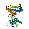
| ||||||||
|---|---|---|---|---|---|---|---|---|---|
| 1 |
| ||||||||
| Unit cell |
|
-Components
| #1: Protein | Mass: 31823.459 Da / Num. of mol.: 1 Source method: isolated from a genetically manipulated source Source: (gene. exp.) Homo sapiens (human) / Gene: EPHB1, ELK, EPHT2, HEK6, NET / Production host:  References: UniProt: P54762, receptor protein-tyrosine kinase |
|---|---|
| #2: Water | ChemComp-HOH /  Mass: 18.015 Da / Num. of mol.: 36 / Source method: isolated from a natural source / Formula: H2O Mass: 18.015 Da / Num. of mol.: 36 / Source method: isolated from a natural source / Formula: H2O |
| Has ligand of interest | Y |
| Has protein modification | Y |
-Experimental details
-Experiment
| Experiment | Method: X-RAY DIFFRACTION / Number of used crystals: 1 |
|---|
- Sample preparation
| Crystal | Density Matthews: 2.5 Å3/Da / Density % sol: 50.77 % |
|---|---|
| Crystal grow | Temperature: 291 K / Method: vapor diffusion, hanging drop / Details: 0.2 M sodium malonate, pH 4.6, 14% PEG3350 |
-Data collection
| Diffraction | Mean temperature: 100 K / Serial crystal experiment: N |
|---|---|
| Diffraction source | Source: SYNCHROTRON / Site: APS  / Beamline: 19-ID / Wavelength: 0.97918 Å / Beamline: 19-ID / Wavelength: 0.97918 Å |
| Detector | Type: DECTRIS PILATUS3 6M / Detector: PIXEL / Date: Nov 2, 2020 |
| Radiation | Monochromator: double crystal Si(111) / Protocol: SINGLE WAVELENGTH / Monochromatic (M) / Laue (L): M / Scattering type: x-ray |
| Radiation wavelength | Wavelength: 0.97918 Å / Relative weight: 1 |
| Reflection | Resolution: 2.7→50 Å / Num. obs: 8602 / % possible obs: 99.7 % / Redundancy: 7.1 % / Rmerge(I) obs: 0.36 / Net I/σ(I): 9.69 |
| Reflection shell | Resolution: 2.7→2.8 Å / Rmerge(I) obs: 1.16 / Num. unique obs: 833 / % possible all: 99.8 |
- Processing
| Software |
| ||||||||||||||||||||||||
|---|---|---|---|---|---|---|---|---|---|---|---|---|---|---|---|---|---|---|---|---|---|---|---|---|---|
| Refinement | Method to determine structure: MOLECULAR REPLACEMENT Starting model: PDB entry 3zfx Resolution: 2.705→48.718 Å / SU ML: 0.31 / Cross valid method: THROUGHOUT / σ(F): 1.35 / Phase error: 23.24 / Stereochemistry target values: ML
| ||||||||||||||||||||||||
| Solvent computation | Shrinkage radii: 0.9 Å / VDW probe radii: 1.11 Å / Solvent model: FLAT BULK SOLVENT MODEL | ||||||||||||||||||||||||
| Displacement parameters | Biso max: 102.24 Å2 / Biso mean: 35.803 Å2 / Biso min: 14.75 Å2 | ||||||||||||||||||||||||
| Refinement step | Cycle: final / Resolution: 2.705→48.718 Å
| ||||||||||||||||||||||||
| LS refinement shell | Refine-ID: X-RAY DIFFRACTION / Rfactor Rfree error: 0
|
