 Movie
Movie Controller

[English] 日本語
 Yorodumi
- PDB-6cfu: Structure of Human alpha-Phosphomannomutase 1 containing mutation... -
+ Open data

- Basic information
| Entry | Database: PDB / ID: 6cfu | ||||||
|---|---|---|---|---|---|---|---|
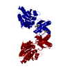
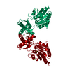
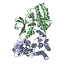
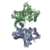
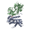
| Title | Structure of Human alpha-Phosphomannomutase 1 containing mutations R180K and R183K | ||||||
 Components Components | Phosphomannomutase 1 | ||||||
 Keywords Keywords | ISOMERASE / phosphatase / haloalkanoate dehalogenase superfamily / mutase | ||||||
| Function / homology |  Function and homology information Function and homology informationSynthesis of GDP-mannose / phosphomannomutase / phosphomannomutase activity / GDP-mannose biosynthetic process / mannose metabolic process / protein N-linked glycosylation / cellular response to leukemia inhibitory factor / neuronal cell body / metal ion binding / cytosol Similarity search - Function | ||||||
| Biological species |  Homo sapiens (human) Homo sapiens (human) | ||||||
| Method |  X-RAY DIFFRACTION / SYNCHROTRON / MOLECULAR REPLACEMENT / Resolution: 2.244 Å X-RAY DIFFRACTION / SYNCHROTRON / MOLECULAR REPLACEMENT / Resolution: 2.244 Å | ||||||
 Authors Authors | Ji, T. / Dunaway-Mariano, D. / Allen, K.N. | ||||||
| Funding support |  United States, 1items United States, 1items
| ||||||
 Citation Citation | Journal: Biochemistry / Year: 2018 Title: Structural Basis of the Molecular Switch between Phosphatase and Mutase Functions of Human Phosphomannomutase 1 under Ischemic Conditions. Authors: Ji, T. / Zhang, C. / Zheng, L. / Dunaway-Mariano, D. / Allen, K.N. | ||||||
| History |
|
- Structure visualization
| Structure viewer | Molecule: MolmilJmol/JSmol |
|---|
- Downloads & links
-Download
| PDBx/mmCIF format | 6cfu.cif.gz | 67.7 KB | Display | PDBx/mmCIF format |
|---|---|---|---|---|
| PDB format | pdb6cfu.ent.gz | 48 KB | Display | PDB format |
| PDBx/mmJSON format | 6cfu.json.gz | Tree view | PDBx/mmJSON format | |
| Others |  Other downloads Other downloads |
-Validation report
| Arichive directory | https://data.pdbj.org/pub/pdb/validation_reports/cf/6cfuftp://data.pdbj.org/pub/pdb/validation_reports/cf/6cfu | HTTPS FTP |
|---|
-Related structure data
| Related structure data |  6cfrC 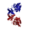 6cfsC 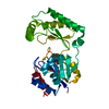 6cftC 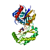 6cfvC  2fucS S: Starting model for refinement C: citing same article ( |
|---|---|
| Similar structure data |


-Links
 PDBj
PDBj- Assembly
| Deposited unit | 
| ||||||||
|---|---|---|---|---|---|---|---|---|---|
| 1 | 
| ||||||||
| Unit cell |
|
-Components
| #1: Protein | Mass: 29730.783 Da / Num. of mol.: 1 / Mutation: R180K, R183K Source method: isolated from a genetically manipulated source Source: (gene. exp.) Homo sapiens (human) / Gene: PMM1, PMMH22 / Production host:  | ||
|---|---|---|---|
| #2: Chemical |   Mass: 24.305 Da / Num. of mol.: 2 / Source method: obtained synthetically / Formula: Mg Mass: 24.305 Da / Num. of mol.: 2 / Source method: obtained synthetically / Formula: Mg#3: Water | ChemComp-HOH / |  Mass: 18.015 Da / Num. of mol.: 123 / Source method: isolated from a natural source / Formula: H2O Mass: 18.015 Da / Num. of mol.: 123 / Source method: isolated from a natural source / Formula: H2O |
-Experimental details
-Experiment
| Experiment | Method: X-RAY DIFFRACTION / Number of used crystals: 1 |
|---|
- Sample preparation
| Crystal | Density Matthews: 2.3 Å3/Da / Density % sol: 46.56 % |
|---|---|
| Crystal grow | Temperature: 290 K / Method: vapor diffusion, hanging drop Details: 20% polyethylene glycol 3350, 0.15 M DL-malate, pH 7.0, 50 mM MgCl2, and 8 mM beta-mercaptoethanol |
-Data collection
| Diffraction | Mean temperature: 100 K |
|---|---|
| Diffraction source | Source: SYNCHROTRON / Site: NSLS / Beamline: X25 / Wavelength: 0.9791 Å |
| Detector | Type: DECTRIS PILATUS 6M / Detector: PIXEL / Date: Aug 21, 2013 |
| Radiation | Protocol: SINGLE WAVELENGTH / Monochromatic (M) / Laue (L): M / Scattering type: x-ray |
| Radiation wavelength | Wavelength: 0.9791 Å / Relative weight: 1 |
| Reflection | Resolution: 2.24→49.28 Å / Num. obs: 13960 / % possible obs: 98.41 % / Redundancy: 2 % / Net I/σ(I): 9.62 |
| Reflection shell | Resolution: 2.24→2.32 Å |
- Processing
| Software |
| |||||||||||||||||||||||||||||||||||||||||||||||||||||||||||||||||||||||||||||
|---|---|---|---|---|---|---|---|---|---|---|---|---|---|---|---|---|---|---|---|---|---|---|---|---|---|---|---|---|---|---|---|---|---|---|---|---|---|---|---|---|---|---|---|---|---|---|---|---|---|---|---|---|---|---|---|---|---|---|---|---|---|---|---|---|---|---|---|---|---|---|---|---|---|---|---|---|---|---|
| Refinement | Method to determine structure: MOLECULAR REPLACEMENT Starting model: 2FUC Resolution: 2.244→49.28 Å / SU ML: 0.3 / Cross valid method: FREE R-VALUE / σ(F): 1.33 / Phase error: 26.72
| |||||||||||||||||||||||||||||||||||||||||||||||||||||||||||||||||||||||||||||
| Solvent computation | Shrinkage radii: 0.9 Å / VDW probe radii: 1.11 Å | |||||||||||||||||||||||||||||||||||||||||||||||||||||||||||||||||||||||||||||
| Refinement step | Cycle: LAST / Resolution: 2.244→49.28 Å
| |||||||||||||||||||||||||||||||||||||||||||||||||||||||||||||||||||||||||||||
| Refine LS restraints |
| |||||||||||||||||||||||||||||||||||||||||||||||||||||||||||||||||||||||||||||
| LS refinement shell |
|
