Synthesis of GDP-mannose / phosphomannomutase / phosphomannomutase activity / GDP-mannose biosynthetic process / mannose metabolic process / protein N-linked glycosylation / cellular response to leukemia inhibitory factor / neuronal cell body / metal ion binding / cytosol Similarity search - Function
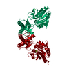
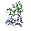
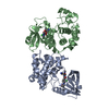
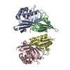
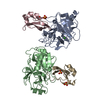
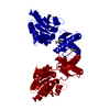
The asymmetric unit contains one monomer of the biologically active dimer. The dimer can be generated by applying the following matrix to the deposited coordinates: | 3.123e-17, 1, 5.881e-16| | 1,-1.745e-16, 1.109e-15| | 1.049e-15, 5.487e-16, -1| (-7.416e-14,-1.349e-13, 216)
-
Components
#1: Protein
Phosphomannomutase1 / PMM 1 / PMMH-22
Mass: 29974.389 Da / Num. of mol.: 1 Source method: isolated from a genetically manipulated source Source: (gene. exp.) Homo sapiens (human) / Plasmid: pET-28b / Production host: Escherichia coli (E. coli) / Strain (production host): BL21(DE3)pLysS / References: UniProt: Q92871, phosphomannomutase
Mass: 18.015 Da / Num. of mol.: 171 / Source method: isolated from a natural source / Formula: H2O
Has protein modification
Y
-
Experimental details
-
Experiment
Experiment
Method: X-RAY DIFFRACTION / Number of used crystals: 1
-
Sample preparation
Crystal
Density Matthews: 2.4 Å3/Da / Density % sol: 48.82 %
Crystal grow
Details: Crystals grown by hanging drop vapor diffusion over mother liquor containing 15-18% polyethylene glycol 3350, 0.15M D,L-malic acid, pH 7.0, 50mM MgCL2, and 8mM 2-mercaptoethanol. Drops were ...Details: Crystals grown by hanging drop vapor diffusion over mother liquor containing 15-18% polyethylene glycol 3350, 0.15M D,L-malic acid, pH 7.0, 50mM MgCL2, and 8mM 2-mercaptoethanol. Drops were formed by mixing 2ul of protein solution (10mM HEPES, pH 7.5, 100mM NaCl, 5mM MgCl2) at 15-25mg/ml with 2ul of mother liquor. Crystals grew after one to three days at 296 K.
In the structure databanks used in Yorodumi, some data are registered as the other names, "COVID-19 virus" and "2019-nCoV". Here are the details of the virus and the list of structure data.
Jan 31, 2019. EMDB accession codes are about to change! (news from PDBe EMDB page)
EMDB accession codes are about to change! (news from PDBe EMDB page)
The allocation of 4 digits for EMDB accession codes will soon come to an end. Whilst these codes will remain in use, new EMDB accession codes will include an additional digit and will expand incrementally as the available range of codes is exhausted. The current 4-digit format prefixed with “EMD-” (i.e. EMD-XXXX) will advance to a 5-digit format (i.e. EMD-XXXXX), and so on. It is currently estimated that the 4-digit codes will be depleted around Spring 2019, at which point the 5-digit format will come into force.
The EM Navigator/Yorodumi systems omit the EMD- prefix.
Related info.:Q: What is EMD? / ID/Accession-code notation in Yorodumi/EM Navigator
Yorodumi is a browser for structure data from EMDB, PDB, SASBDB, etc.
This page is also the successor to EM Navigator detail page, and also detail information page/front-end page for Omokage search.
The word "yorodu" (or yorozu) is an old Japanese word meaning "ten thousand". "mi" (miru) is to see.
Related info.:EMDB / PDB / SASBDB / Comparison of 3 databanks / Yorodumi Search / Aug 31, 2016. New EM Navigator & Yorodumi / Yorodumi Papers / Jmol/JSmol / Function and homology information / Changes in new EM Navigator and Yorodumi
 Movie
Movie Controller


 Open data
 Basic information
 Components
 Keywords
 Function and homology information
 Homo sapiens (human)
 X-RAY DIFFRACTION /
  Authors
 Citation
 Structure visualization
 Downloads & links
 Other downloads
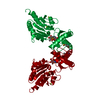


 PDBj
PDBj Assembly
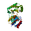


 Mass: 24.305 Da / Num. of mol.: 2 / Source method: obtained synthetically / Formula: Mg
 Mass: 18.015 Da / Num. of mol.: 171 / Source method: isolated from a natural source / Formula: H2O
 Sample preparation
 / Beamline: X12C / Wavelength: 0.9797, 0.9794, 0.9500
 Processing