 Movie
Movie Controller

[English] 日本語
 Yorodumi
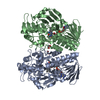
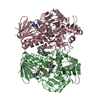
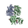
- PDB-6fpj: Structure of the AMPAR GluA3 N-terminal domain bound to phosphate -
+ Open data

- Basic information
| Entry | Database: PDB / ID: 6fpj | ||||||
|---|---|---|---|---|---|---|---|
| Title | Structure of the AMPAR GluA3 N-terminal domain bound to phosphate | ||||||
 Components Components | Glutamate receptor 3 | ||||||
 Keywords Keywords | MEMBRANE PROTEIN / ligand-gated ion channel / AMPA receptor | ||||||
| Function / homology |  Function and homology information Function and homology informationTrafficking of AMPA receptors / ligand-gated calcium channel activity / Synaptic adhesion-like molecules / Activation of AMPA receptors / protein heterotetramerization / Trafficking of GluR2-containing AMPA receptors / response to lithium ion / AMPA glutamate receptor activity / parallel fiber to Purkinje cell synapse / AMPA glutamate receptor complex ...Trafficking of AMPA receptors / ligand-gated calcium channel activity / Synaptic adhesion-like molecules / Activation of AMPA receptors / protein heterotetramerization / Trafficking of GluR2-containing AMPA receptors / response to lithium ion / AMPA glutamate receptor activity / parallel fiber to Purkinje cell synapse / AMPA glutamate receptor complex / regulation of receptor recycling / cellular response to glycine / ionotropic glutamate receptor complex / asymmetric synapse / Unblocking of NMDA receptors, glutamate binding and activation / synaptic cleft / response to fungicide / glutamate-gated receptor activity / glutamate-gated calcium ion channel activity / presynaptic active zone membrane / ligand-gated monoatomic ion channel activity involved in regulation of presynaptic membrane potential / dendritic shaft / synaptic transmission, glutamatergic / transmitter-gated monoatomic ion channel activity involved in regulation of postsynaptic membrane potential / postsynaptic density membrane / modulation of chemical synaptic transmission / terminal bouton / long-term synaptic potentiation / amyloid-beta binding / presynaptic membrane / protein homotetramerization / dendritic spine / perikaryon / postsynaptic membrane / postsynaptic density / neuronal cell body / dendrite / glutamatergic synapse / protein-containing complex / membrane / plasma membrane Similarity search - Function | ||||||
| Biological species |  | ||||||
| Method |  X-RAY DIFFRACTION / SYNCHROTRON / MOLECULAR REPLACEMENT / Resolution: 1.96 Å X-RAY DIFFRACTION / SYNCHROTRON / MOLECULAR REPLACEMENT / Resolution: 1.96 Å | ||||||
 Authors Authors | Herguedas, B. / Garcia-Nafria, J. / Greger, I. | ||||||
 Citation Citation | Journal: Structure / Year: 2019 Title: Druggability Simulations and X-Ray Crystallography Reveal a Ligand-Binding Site in the GluA3 AMPA Receptor N-Terminal Domain. Authors: Lee, J.Y. / Krieger, J. / Herguedas, B. / Garcia-Nafria, J. / Dutta, A. / Shaikh, S.A. / Greger, I.H. / Bahar, I. | ||||||
| History |
|
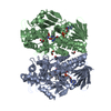
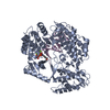
- Structure visualization
| Structure viewer | Molecule: MolmilJmol/JSmol |
|---|
- Downloads & links
-Download
| PDBx/mmCIF format | 6fpj.cif.gz | 471.6 KB | Display | PDBx/mmCIF format |
|---|---|---|---|---|
| PDB format | pdb6fpj.ent.gz | 389.3 KB | Display | PDB format |
| PDBx/mmJSON format | 6fpj.json.gz | Tree view | PDBx/mmJSON format | |
| Others |  Other downloads Other downloads |
-Validation report
| Arichive directory | https://data.pdbj.org/pub/pdb/validation_reports/fp/6fpjftp://data.pdbj.org/pub/pdb/validation_reports/fp/6fpj | HTTPS FTP |
|---|
-Related structure data
| Related structure data |  6flrC 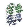 3o21S C: citing same article ( S: Starting model for refinement |
|---|---|
| Similar structure data |

-Links
 PDBj
PDBj


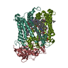
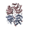
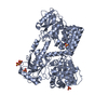
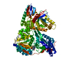
- Assembly
| Deposited unit | 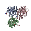
| |||||||||||||||||||||||||||||||||||||||||||||||||||||
|---|---|---|---|---|---|---|---|---|---|---|---|---|---|---|---|---|---|---|---|---|---|---|---|---|---|---|---|---|---|---|---|---|---|---|---|---|---|---|---|---|---|---|---|---|---|---|---|---|---|---|---|---|---|---|
| 1 | 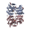
| |||||||||||||||||||||||||||||||||||||||||||||||||||||
| 2 | 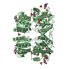
| |||||||||||||||||||||||||||||||||||||||||||||||||||||
| Unit cell |
| |||||||||||||||||||||||||||||||||||||||||||||||||||||
| Components on special symmetry positions |
| |||||||||||||||||||||||||||||||||||||||||||||||||||||
| Noncrystallographic symmetry (NCS) | NCS domain:
NCS domain segments: Component-ID: _ / Beg auth comp-ID: PHE / Beg label comp-ID: PHE / End auth comp-ID: PHE / End label comp-ID: PHE / Refine code: _ / Auth seq-ID: 2 - 380 / Label seq-ID: 2 - 380
NCS ensembles :
|
-Components
-Protein / Sugars , 2 types, 11 molecules ABC
| #1: Protein | Mass: 45093.953 Da / Num. of mol.: 3 Source method: isolated from a genetically manipulated source Source: (gene. exp.)  Homo sapiens (human) / References: UniProt: P19492 Homo sapiens (human) / References: UniProt: P19492#2: Sugar | ChemComp-NAG /  Type: D-saccharide, beta linking / Mass: 221.208 Da / Num. of mol.: 8 Type: D-saccharide, beta linking / Mass: 221.208 Da / Num. of mol.: 8Source method: isolated from a genetically manipulated source Formula: C8H15NO6 |
|---|
-Non-polymers , 4 types, 668 molecules 


| #3: Chemical | ChemComp-PO4 /  Mass: 94.971 Da / Num. of mol.: 7 Mass: 94.971 Da / Num. of mol.: 7Source method: isolated from a genetically manipulated source Formula: PO4 #4: Chemical | ChemComp-DMS /  Mass: 78.133 Da / Num. of mol.: 7 / Source method: obtained synthetically / Formula: C2H6OS / Comment: DMSO, precipitant*YM Mass: 78.133 Da / Num. of mol.: 7 / Source method: obtained synthetically / Formula: C2H6OS / Comment: DMSO, precipitant*YM#5: Chemical | ChemComp-GOL / |  Mass: 92.094 Da / Num. of mol.: 1 / Source method: obtained synthetically / Formula: C3H8O3 Mass: 92.094 Da / Num. of mol.: 1 / Source method: obtained synthetically / Formula: C3H8O3#6: Water | ChemComp-HOH / | Mass: 18.015 Da / Num. of mol.: 653 / Source method: isolated from a natural source / Formula: H2O |
|---|
-Details
| Has protein modification | Y |
|---|
-Experimental details
-Experiment
| Experiment | Method: X-RAY DIFFRACTION / Number of used crystals: 1 |
|---|
- Sample preparation
| Crystal | Density Matthews: 2.17 Å3/Da / Density % sol: 43.23 % |
|---|---|
| Crystal grow | Temperature: 293 K / Method: vapor diffusion / Details: Ammonium dihydrogen phosphate pH 4.6 PEG3350 |
-Data collection
| Diffraction | Mean temperature: 100 K |
|---|---|
| Diffraction source | Source: SYNCHROTRON / Site: Diamond  / Beamline: I04 / Wavelength: 0.9 Å / Beamline: I04 / Wavelength: 0.9 Å |
| Detector | Type: ADSC QUANTUM 315 / Detector: CCD / Date: Oct 31, 2011 |
| Radiation | Protocol: SINGLE WAVELENGTH / Monochromatic (M) / Laue (L): M / Scattering type: x-ray |
| Radiation wavelength | Wavelength: 0.9 Å / Relative weight: 1 |
| Reflection | Resolution: 1.96→164.77 Å / Num. obs: 81888 / % possible obs: 98.9 % / Redundancy: 3.8 % / Rmerge(I) obs: 0.063 / Net I/σ(I): 12.9 |
| Reflection shell | Resolution: 1.96→2.01 Å |
- Processing
| Software |
| ||||||||||||||||||||||||||||||||||||||||||||||||||||||||||||||||||||||||||||||||||||||||||||||||||||
|---|---|---|---|---|---|---|---|---|---|---|---|---|---|---|---|---|---|---|---|---|---|---|---|---|---|---|---|---|---|---|---|---|---|---|---|---|---|---|---|---|---|---|---|---|---|---|---|---|---|---|---|---|---|---|---|---|---|---|---|---|---|---|---|---|---|---|---|---|---|---|---|---|---|---|---|---|---|---|---|---|---|---|---|---|---|---|---|---|---|---|---|---|---|---|---|---|---|---|---|---|---|
| Refinement | Method to determine structure: MOLECULAR REPLACEMENT Starting model: 3O21 Resolution: 1.96→91 Å / Cor.coef. Fo:Fc: 0.956 / Cor.coef. Fo:Fc free: 0.934 / SU B: 8.097 / SU ML: 0.117 / Cross valid method: THROUGHOUT / σ(F): 0 / ESU R: 0.186 / ESU R Free: 0.158 / Stereochemistry target values: MAXIMUM LIKELIHOOD Details: HYDROGENS HAVE BEEN ADDED IN THE RIDING POSITIONS U VALUES : WITH TLS ADDED
| ||||||||||||||||||||||||||||||||||||||||||||||||||||||||||||||||||||||||||||||||||||||||||||||||||||
| Solvent computation | Ion probe radii: 0.8 Å / Shrinkage radii: 0.8 Å / VDW probe radii: 1.2 Å / Solvent model: MASK | ||||||||||||||||||||||||||||||||||||||||||||||||||||||||||||||||||||||||||||||||||||||||||||||||||||
| Displacement parameters | Biso max: 88.6 Å2 / Biso mean: 28.181 Å2 / Biso min: 10.89 Å2
| ||||||||||||||||||||||||||||||||||||||||||||||||||||||||||||||||||||||||||||||||||||||||||||||||||||
| Refinement step | Cycle: final / Resolution: 1.96→91 Å
| ||||||||||||||||||||||||||||||||||||||||||||||||||||||||||||||||||||||||||||||||||||||||||||||||||||
| Refine LS restraints |
| ||||||||||||||||||||||||||||||||||||||||||||||||||||||||||||||||||||||||||||||||||||||||||||||||||||
| Refine LS restraints NCS | Refine-ID: X-RAY DIFFRACTION / Type: interatomic distance / Weight position: 0.05
| ||||||||||||||||||||||||||||||||||||||||||||||||||||||||||||||||||||||||||||||||||||||||||||||||||||
| LS refinement shell | Resolution: 1.96→2.011 Å / Rfactor Rfree error: 0 / Total num. of bins used: 20
| ||||||||||||||||||||||||||||||||||||||||||||||||||||||||||||||||||||||||||||||||||||||||||||||||||||
| Refinement TLS params. | Method: refined / Refine-ID: X-RAY DIFFRACTION
| ||||||||||||||||||||||||||||||||||||||||||||||||||||||||||||||||||||||||||||||||||||||||||||||||||||
| Refinement TLS group |
|
