 Movie
Movie Controller

[English] 日本語
 Yorodumi
- PDB-7jfp: GTP-specific succinyl-CoA synthetase complexed with Mg-GDP, phosp... -
+ Open data

- Basic information
| Entry | Database: PDB / ID: 7jfp | ||||||
|---|---|---|---|---|---|---|---|
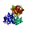
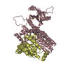
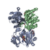
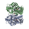
| Title | GTP-specific succinyl-CoA synthetase complexed with Mg-GDP, phosphohistidine loop pointing towards nucleotide binding site | ||||||
 Components Components |
| ||||||
 Keywords Keywords | LIGASE / Complex | ||||||
| Function / homology |  Function and homology information Function and homology informationmalate-CoA ligase / succinate-CoA ligase complex (GDP-forming) / succinate-CoA ligase (GDP-forming) / succinate-CoA ligase (GDP-forming) activity / succinate-CoA ligase complex (ADP-forming) / Citric acid cycle (TCA cycle) / succinate-CoA ligase (ADP-forming) / succinate-CoA ligase complex / Ligases; Forming carbon-sulfur bonds; Acid-thiol ligases / succinate-CoA ligase (ADP-forming) activity ...malate-CoA ligase / succinate-CoA ligase complex (GDP-forming) / succinate-CoA ligase (GDP-forming) / succinate-CoA ligase (GDP-forming) activity / succinate-CoA ligase complex (ADP-forming) / Citric acid cycle (TCA cycle) / succinate-CoA ligase (ADP-forming) / succinate-CoA ligase complex / Ligases; Forming carbon-sulfur bonds; Acid-thiol ligases / succinate-CoA ligase (ADP-forming) activity / succinyl-CoA metabolic process / tricarboxylic acid cycle / nucleotide binding / GTP binding / mitochondrion / ATP binding / metal ion binding Similarity search - Function | ||||||
| Biological species |  | ||||||
| Method |  X-RAY DIFFRACTION / SYNCHROTRON / MOLECULAR REPLACEMENT / Resolution: 2.55 Å X-RAY DIFFRACTION / SYNCHROTRON / MOLECULAR REPLACEMENT / Resolution: 2.55 Å | ||||||
 Authors Authors | Huang, J. / Fraser, M.E. | ||||||
| Funding support |  Canada, 1items Canada, 1items
| ||||||
 Citation Citation | Journal: Acta Crystallogr.,Sect.D / Year: 2021 Title: Second distinct conformation of the phosphohistidine loop in succinyl-CoA synthetase Authors: Huang, J. / Fraser, M.E. | ||||||
| History |
|
- Structure visualization
| Structure viewer | Molecule: MolmilJmol/JSmol |
|---|
- Downloads & links
-Download
| PDBx/mmCIF format | 7jfp.cif.gz | 400.1 KB | Display | PDBx/mmCIF format |
|---|---|---|---|---|
| PDB format | pdb7jfp.ent.gz | 331.5 KB | Display | PDB format |
| PDBx/mmJSON format | 7jfp.json.gz | Tree view | PDBx/mmJSON format | |
| Others |  Other downloads Other downloads |
-Validation report
| Arichive directory | https://data.pdbj.org/pub/pdb/validation_reports/jf/7jfpftp://data.pdbj.org/pub/pdb/validation_reports/jf/7jfp | HTTPS FTP |
|---|
-Related structure data
| Related structure data | 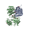 6xruC 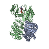 7jj0C 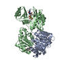 7jkrC 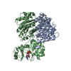 7jmkC 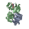 2fp4S S: Starting model for refinement C: citing same article ( |
|---|---|
| Similar structure data |


-Links
 PDBj
PDBj


- Assembly
| Deposited unit | 
| ||||||||
|---|---|---|---|---|---|---|---|---|---|
| 1 |
| ||||||||
| Unit cell |
|
-Components
-Protein , 2 types, 2 molecules AB
| #1: Protein | Mass: 32089.859 Da / Num. of mol.: 1 Source method: isolated from a genetically manipulated source Source: (gene. exp.)  References: UniProt: O19069, succinate-CoA ligase (GDP-forming), succinate-CoA ligase (ADP-forming) |
|---|---|
| #2: Protein | Mass: 42711.867 Da / Num. of mol.: 1 Source method: isolated from a genetically manipulated source Source: (gene. exp.) References: UniProt: P53590, succinate-CoA ligase (GDP-forming) |
-Non-polymers , 4 types, 11 molecules 


| #3: Chemical |  Mass: 92.094 Da / Num. of mol.: 3 / Source method: obtained synthetically / Formula: C3H8O3 Mass: 92.094 Da / Num. of mol.: 3 / Source method: obtained synthetically / Formula: C3H8O3#4: Chemical | ChemComp-GDP / |  Type: RNA linking / Mass: 443.201 Da / Num. of mol.: 1 / Source method: obtained synthetically / Formula: C10H15N5O11P2 / Feature type: SUBJECT OF INVESTIGATION / Comment: GDP, energy-carrying molecule*YM Type: RNA linking / Mass: 443.201 Da / Num. of mol.: 1 / Source method: obtained synthetically / Formula: C10H15N5O11P2 / Feature type: SUBJECT OF INVESTIGATION / Comment: GDP, energy-carrying molecule*YM#5: Chemical | ChemComp-MG / |  Mass: 24.305 Da / Num. of mol.: 1 / Source method: obtained synthetically / Formula: Mg Mass: 24.305 Da / Num. of mol.: 1 / Source method: obtained synthetically / Formula: Mg#6: Water | ChemComp-HOH / | Mass: 18.015 Da / Num. of mol.: 6 / Source method: isolated from a natural source / Formula: H2O |
|---|
-Details
| Has ligand of interest | Y |
|---|---|
| Has protein modification | Y |
-Experimental details
-Experiment
| Experiment | Method: X-RAY DIFFRACTION / Number of used crystals: 1 |
|---|
- Sample preparation
| Crystal | Density Matthews: 2.57 Å3/Da / Density % sol: 52.18 % |
|---|---|
| Crystal grow | Temperature: 294 K / Method: vapor diffusion, hanging drop / Details: 1.0 M sodium citrate, Tris-HCl pH 7.4 |
-Data collection
| Diffraction | Mean temperature: 100 K / Serial crystal experiment: N |
|---|---|
| Diffraction source | Source: SYNCHROTRON / Site: ALS  / Beamline: 5.0.2 / Wavelength: 1 Å / Beamline: 5.0.2 / Wavelength: 1 Å |
| Detector | Type: DECTRIS PILATUS3 6M / Detector: PIXEL / Date: Jun 1, 2019 |
| Radiation | Protocol: SINGLE WAVELENGTH / Monochromatic (M) / Laue (L): M / Scattering type: x-ray |
| Radiation wavelength | Wavelength: 1 Å / Relative weight: 1 |
| Reflection | Resolution: 2.55→52.99 Å / Num. obs: 25660 / % possible obs: 99.8 % / Redundancy: 4.6 % / CC1/2: 0.999 / Rmerge(I) obs: 0.084 / Rpim(I) all: 0.063 / Net I/σ(I): 8.1 |
| Reflection shell | Resolution: 2.55→2.59 Å / Mean I/σ(I) obs: 0.2 / Num. unique obs: 1278 / CC1/2: 0.297 |
- Processing
| Software |
| |||||||||||||||||||||||||||||||||||||||||||||||||||||||||||||||
|---|---|---|---|---|---|---|---|---|---|---|---|---|---|---|---|---|---|---|---|---|---|---|---|---|---|---|---|---|---|---|---|---|---|---|---|---|---|---|---|---|---|---|---|---|---|---|---|---|---|---|---|---|---|---|---|---|---|---|---|---|---|---|---|---|
| Refinement | Method to determine structure: MOLECULAR REPLACEMENT Starting model: 2FP4 Resolution: 2.55→52.99 Å / SU ML: 0.49 / Cross valid method: THROUGHOUT / σ(F): 1.33 / Phase error: 34.69 / Stereochemistry target values: ML
| |||||||||||||||||||||||||||||||||||||||||||||||||||||||||||||||
| Solvent computation | Shrinkage radii: 0.9 Å / VDW probe radii: 1.11 Å / Solvent model: FLAT BULK SOLVENT MODEL | |||||||||||||||||||||||||||||||||||||||||||||||||||||||||||||||
| Displacement parameters | Biso max: 283.58 Å2 / Biso mean: 113.4011 Å2 / Biso min: 48.63 Å2 | |||||||||||||||||||||||||||||||||||||||||||||||||||||||||||||||
| Refinement step | Cycle: final / Resolution: 2.55→52.99 Å
| |||||||||||||||||||||||||||||||||||||||||||||||||||||||||||||||
| LS refinement shell | Refine-ID: X-RAY DIFFRACTION / Rfactor Rfree error: 0 / Total num. of bins used: 8
| |||||||||||||||||||||||||||||||||||||||||||||||||||||||||||||||
| Refinement TLS params. | Method: refined / Origin x: -7.3836 Å / Origin y: 28.0442 Å / Origin z: 13.0689 Å
| |||||||||||||||||||||||||||||||||||||||||||||||||||||||||||||||
| Refinement TLS group |
|
