 Movie
Movie Controller


+ Open data

- Basic information
| Entry | Database: PDB / ID: 6fh2 | ||||||
|---|---|---|---|---|---|---|---|
| Title | Protein arginine kinase McsB in the AMP-PN-bound state | ||||||
 Components Components | Protein-arginine kinase | ||||||
 Keywords Keywords | SIGNALING PROTEIN / protein kinase / protein arginine kinase / protein arginine phosphorylation / phospho-binding domain | ||||||
| Function / homology |  Function and homology information Function and homology informationprotein arginine kinase / protein arginine kinase activity / phosphocreatine biosynthetic process / creatine kinase activity / : / ATP binding Similarity search - Function | ||||||
| Biological species |   Geobacillus stearothermophilus (bacteria) Geobacillus stearothermophilus (bacteria) | ||||||
| Method |  X-RAY DIFFRACTION / SYNCHROTRON / MOLECULAR REPLACEMENT / Resolution: 2.7 Å X-RAY DIFFRACTION / SYNCHROTRON / MOLECULAR REPLACEMENT / Resolution: 2.7 Å | ||||||
 Authors Authors | Suskiewicz, M.J. / Heuck, A. / Vu, L.D. / Clausen, T. | ||||||
 Citation Citation | Journal: Nat.Chem.Biol. / Year: 2019 Title: Structure of McsB, a protein kinase for regulated arginine phosphorylation. Authors: Suskiewicz, M.J. / Hajdusits, B. / Beveridge, R. / Heuck, A. / Vu, L.D. / Kurzbauer, R. / Hauer, K. / Thoeny, V. / Rumpel, K. / Mechtler, K. / Meinhart, A. / Clausen, T. | ||||||
| History |
|
- Structure visualization
| Structure viewer | Molecule: MolmilJmol/JSmol |
|---|
- Downloads & links
-Download
| PDBx/mmCIF format | 6fh2.cif.gz | 144.8 KB | Display | PDBx/mmCIF format |
|---|---|---|---|---|
| PDB format | pdb6fh2.ent.gz | 112.5 KB | Display | PDB format |
| PDBx/mmJSON format | 6fh2.json.gz | Tree view | PDBx/mmJSON format | |
| Others |  Other downloads Other downloads |
-Validation report
| Arichive directory | https://data.pdbj.org/pub/pdb/validation_reports/fh/6fh2ftp://data.pdbj.org/pub/pdb/validation_reports/fh/6fh2 | HTTPS FTP |
|---|
-Related structure data
| Related structure data | 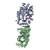 6fh1C 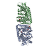 6fh3C 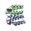 6fh4C 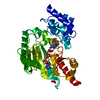 1m15S C: citing same article ( S: Starting model for refinement |
|---|---|
| Similar structure data |


-Links
 PDBj
PDBj
- Assembly
| Deposited unit | 
| ||||||||
|---|---|---|---|---|---|---|---|---|---|
| 1 |
| ||||||||
| Unit cell |
|
-Components
| #1: Protein | Mass: 41255.207 Da / Num. of mol.: 2 Source method: isolated from a genetically manipulated source Source: (gene. exp.) Geobacillus stearothermophilus (bacteria)Gene: mcsB / Plasmid: pET-21a(+) / Production host: #2: Chemical | ChemComp-AN2 / |   Mass: 426.216 Da / Num. of mol.: 1 / Source method: obtained synthetically / Formula: C10H16N6O9P2 Mass: 426.216 Da / Num. of mol.: 1 / Source method: obtained synthetically / Formula: C10H16N6O9P2#3: Chemical | ChemComp-EDO /   Mass: 62.068 Da / Num. of mol.: 6 / Source method: isolated from a natural source / Formula: C2H6O2 Mass: 62.068 Da / Num. of mol.: 6 / Source method: isolated from a natural source / Formula: C2H6O2#4: Water | ChemComp-HOH / |  Mass: 18.015 Da / Num. of mol.: 33 / Source method: isolated from a natural source / Formula: H2O Mass: 18.015 Da / Num. of mol.: 33 / Source method: isolated from a natural source / Formula: H2O |
|---|
-Experimental details
-Experiment
| Experiment | Method: X-RAY DIFFRACTION / Number of used crystals: 1 |
|---|
- Sample preparation
| Crystal | Density Matthews: 2.36 Å3/Da / Density % sol: 47.9 % |
|---|---|
| Crystal grow | Temperature: 277.15 K / Method: vapor diffusion, sitting drop / pH: 6.2 Details: 25% v/v ethylene glycol, 75 mM MES-imidazole, 58 mM sodium formate, 58 mM ammonium acetate, 58 mM sodium citrate, 58 mM sodium potassium tartrate, 58 mM sodium oxamate. Soaked with 10 mM ...Details: 25% v/v ethylene glycol, 75 mM MES-imidazole, 58 mM sodium formate, 58 mM ammonium acetate, 58 mM sodium citrate, 58 mM sodium potassium tartrate, 58 mM sodium oxamate. Soaked with 10 mM magnesium AMPPNP, which apparently hydrolysed during the procedure to ADP. |
-Data collection
| Diffraction | Mean temperature: 100 K / Serial crystal experiment: N |
|---|---|
| Diffraction source | Source: SYNCHROTRON / Site: ESRF  / Beamline: ID23-2 / Wavelength: 0.873 Å / Beamline: ID23-2 / Wavelength: 0.873 Å |
| Detector | Type: DECTRIS PILATUS3 2M / Detector: PIXEL / Date: Mar 10, 2014 |
| Radiation | Protocol: SINGLE WAVELENGTH / Monochromatic (M) / Laue (L): M / Scattering type: x-ray |
| Radiation wavelength | Wavelength: 0.873 Å / Relative weight: 1 |
| Reflection | Resolution: 2.7→48 Å / Num. obs: 21210 / % possible obs: 99.76 % / Redundancy: 11.1 % / Biso Wilson estimate: 62.63 Å2 / Net I/σ(I): 15.99 |
| Reflection shell | Resolution: 2.7→2.8 Å |
- Processing
| Software |
| |||||||||||||||||||||||||||||||||||||||||||||||||||||||||||||||||||||||||||||||||||||||||||||||||||||||||
|---|---|---|---|---|---|---|---|---|---|---|---|---|---|---|---|---|---|---|---|---|---|---|---|---|---|---|---|---|---|---|---|---|---|---|---|---|---|---|---|---|---|---|---|---|---|---|---|---|---|---|---|---|---|---|---|---|---|---|---|---|---|---|---|---|---|---|---|---|---|---|---|---|---|---|---|---|---|---|---|---|---|---|---|---|---|---|---|---|---|---|---|---|---|---|---|---|---|---|---|---|---|---|---|---|---|---|
| Refinement | Method to determine structure: MOLECULAR REPLACEMENT Starting model: 1M15 Resolution: 2.7→47.999 Å / SU ML: 0.36 / Cross valid method: FREE R-VALUE / σ(F): 1.37 / Phase error: 31.61 / Stereochemistry target values: ML
| |||||||||||||||||||||||||||||||||||||||||||||||||||||||||||||||||||||||||||||||||||||||||||||||||||||||||
| Solvent computation | Shrinkage radii: 0.9 Å / VDW probe radii: 1.11 Å / Solvent model: FLAT BULK SOLVENT MODEL | |||||||||||||||||||||||||||||||||||||||||||||||||||||||||||||||||||||||||||||||||||||||||||||||||||||||||
| Refinement step | Cycle: LAST / Resolution: 2.7→47.999 Å
| |||||||||||||||||||||||||||||||||||||||||||||||||||||||||||||||||||||||||||||||||||||||||||||||||||||||||
| Refine LS restraints |
| |||||||||||||||||||||||||||||||||||||||||||||||||||||||||||||||||||||||||||||||||||||||||||||||||||||||||
| LS refinement shell |
|
