 Movie
Movie Controller


+ Open data

- Basic information
| Entry | Database: PDB / ID: 1m15 | ||||||
|---|---|---|---|---|---|---|---|
| Title | Transition state structure of arginine kinase | ||||||
 Components Components | arginine kinase | ||||||
 Keywords Keywords | TRANSFERASE / ARGININE KINASE / CREATINE KINASE / PHOSPHAGEN KINASE / TRANSITION STATE ANALOG / ADENOSINE TRIPHOSPHATE | ||||||
| Function / homology |  Function and homology information Function and homology informationarginine kinase / arginine kinase activity / phosphocreatine biosynthetic process / creatine kinase activity / phosphorylation / : / ATP binding / cytoplasm Similarity search - Function | ||||||
| Biological species |  Limulus polyphemus (Atlantic horseshoe crab) Limulus polyphemus (Atlantic horseshoe crab) | ||||||
| Method |  X-RAY DIFFRACTION / SYNCHROTRON / MOLECULAR REPLACEMENT / Resolution: 1.2 Å X-RAY DIFFRACTION / SYNCHROTRON / MOLECULAR REPLACEMENT / Resolution: 1.2 Å | ||||||
 Authors Authors | Yousef, M.S. / Fabiola, F. / Gattis, J.L. / Somasundaram, T. / Chapman, M.S. | ||||||
 Citation Citation | Journal: Acta Crystallogr.,Sect.D / Year: 2002 Title: Refinement of the arginine kinase transition-state analogue complex at 1.2 A resolution: mechanistic insights. Authors: Yousef, M.S. / Fabiola, F. / Gattis, J.L. / Somasundaram, T. / Chapman, M.S. #1: Journal: Acta Crystallogr.,Sect.D / Year: 1999Title: Critical Initial Real-Space Refinement in the Structure Determination of Arginine Kinase Authors: Zhou, G. / Somasundaram, T. / Blanc, E. / Chen, Z. / Chapman, M.S. #2: Journal: Protein Sci. / Year: 1997Title: Expression, Purification from Inclusion Bodies, and Crystal Characterization of a Transition State Analog Complex of Arginine Kinase: A Model for Studying Phosphagen Kinases Authors: Zhou, G. / Parthasarathy, G. / Somasundaram, T. / Ables, A. / Roy, L. / Strong, S.J. / Ellington, W.R. / Chapman, M.S. #3: Journal: Proc.Natl.Acad.Sci.USA / Year: 1998Title: Transition State Structure of Arginine Kinase: Implications for Catalysis of Bimolecular Reactions Authors: Zhou, G. / Somasundaram, T. / Blanc, E. / Parthasarathy, G. / Ellington, W.R. / Chapman, M.S. | ||||||
| History |
|
- Structure visualization
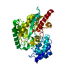
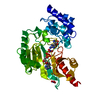
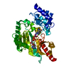
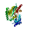
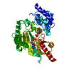
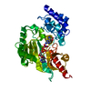
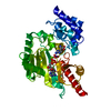
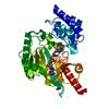
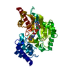
| Structure viewer | Molecule: MolmilJmol/JSmol |
|---|
- Downloads & links
-Download
| PDBx/mmCIF format | 1m15.cif.gz | 182.9 KB | Display | PDBx/mmCIF format |
|---|---|---|---|---|
| PDB format | pdb1m15.ent.gz | 143.4 KB | Display | PDB format |
| PDBx/mmJSON format | 1m15.json.gz | Tree view | PDBx/mmJSON format | |
| Others |  Other downloads Other downloads |
-Validation report
| Arichive directory | https://data.pdbj.org/pub/pdb/validation_reports/m1/1m15ftp://data.pdbj.org/pub/pdb/validation_reports/m1/1m15 | HTTPS FTP |
|---|
-Related structure data
| Related structure data | 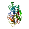 1bgoS S: Starting model for refinement |
|---|---|
| Similar structure data |

-Links
 PDBj
PDBj

- Assembly
| Deposited unit | 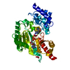
| ||||||||
|---|---|---|---|---|---|---|---|---|---|
| 1 |
| ||||||||
| Unit cell |
|
-Components
-Protein , 1 types, 1 molecules A
| #1: Protein | Mass: 40249.758 Da / Num. of mol.: 1 / Mutation: E103Q, D112G, G116A Source method: isolated from a genetically manipulated source Source: (gene. exp.) Limulus polyphemus (Atlantic horseshoe crab)Plasmid: pET22b / Production host:  |
|---|
-Non-polymers , 5 types, 562 molecules 


| #2: Chemical |  Mass: 62.005 Da / Num. of mol.: 2 / Source method: obtained synthetically / Formula: NO3 Mass: 62.005 Da / Num. of mol.: 2 / Source method: obtained synthetically / Formula: NO3#3: Chemical | ChemComp-MG / |  Mass: 24.305 Da / Num. of mol.: 1 / Source method: obtained synthetically / Formula: Mg Mass: 24.305 Da / Num. of mol.: 1 / Source method: obtained synthetically / Formula: Mg#4: Chemical | ChemComp-ARG / |  Type: L-peptide linking / Mass: 175.209 Da / Num. of mol.: 1 / Source method: obtained synthetically / Formula: C6H15N4O2 Type: L-peptide linking / Mass: 175.209 Da / Num. of mol.: 1 / Source method: obtained synthetically / Formula: C6H15N4O2#5: Chemical | ChemComp-ADP / |  Mass: 427.201 Da / Num. of mol.: 1 / Source method: obtained synthetically / Formula: C10H15N5O10P2 / Comment: ADP, energy-carrying molecule*YM Mass: 427.201 Da / Num. of mol.: 1 / Source method: obtained synthetically / Formula: C10H15N5O10P2 / Comment: ADP, energy-carrying molecule*YM#6: Water | ChemComp-HOH / | Mass: 18.015 Da / Num. of mol.: 557 / Source method: isolated from a natural source / Formula: H2O |
|---|
-Experimental details
-Experiment
| Experiment | Method: X-RAY DIFFRACTION / Number of used crystals: 1 |
|---|
- Sample preparation
| Crystal | Density Matthews: 1.98 Å3/Da / Density % sol: 46.36 % |
|---|---|
| Crystal grow | pH: 8 / Details: pH 8 |
| Crystal grow | *PLUS pH: 7.5 / Method: vapor diffusion, hanging drop / Details: Zhou, G., (1997) Protein Sci., 6, 444. |
| Components of the solutions | *PLUS Conc.: 15-20 % / Common name: PEG6000 / Details: pH7.5 |
-Data collection
| Diffraction | Mean temperature: 100 K |
|---|---|
| Diffraction source | Source: SYNCHROTRON / Site: NSLS  / Beamline: X12B / Wavelength: 0.95 / Beamline: X12B / Wavelength: 0.95 |
| Detector | Type: ADSC QUANTUM 4 / Detector: CCD / Date: Apr 22, 1999 |
| Radiation | Monochromator: DOUBLE CRYSTAL / Protocol: SINGLE WAVELENGTH / Monochromatic (M) / Laue (L): M / Scattering type: x-ray |
| Radiation wavelength | Wavelength: 0.95 Å / Relative weight: 1 |
| Reflection | Resolution: 1.2→20 Å / Num. obs: 115910 / % possible obs: 96.2 % / Observed criterion σ(I): 0 / Rsym value: 0.048 / Net I/σ(I): 24 |
| Reflection shell | Resolution: 1.2→1.23 Å / Mean I/σ(I) obs: 2.1 / Rsym value: 0.323 / % possible all: 71.5 |
| Reflection | *PLUS Lowest resolution: 20 Å / Num. measured all: 1112071 / Rmerge(I) obs: 0.048 |
| Reflection shell | *PLUS Highest resolution: 1.2 Å / % possible obs: 71.5 % / Rmerge(I) obs: 0.323 |
- Processing
| Software |
| |||||||||||||||||||||||||||||||||
|---|---|---|---|---|---|---|---|---|---|---|---|---|---|---|---|---|---|---|---|---|---|---|---|---|---|---|---|---|---|---|---|---|---|---|
| Refinement | Method to determine structure: MOLECULAR REPLACEMENT Starting model: PDB ENTRY 1BGO Resolution: 1.2→6 Å / Num. parameters: 31135 / Num. restraintsaints: 37290 / Cross valid method: THROUGHOUT / σ(F): 0 / Stereochemistry target values: ENGH AND HUBER Details: ANISOTROPIC REFINEMENT REDUCED FREE R (NO CUTOFF) BY 4%
| |||||||||||||||||||||||||||||||||
| Refine analyze | Num. disordered residues: 8 / Occupancy sum hydrogen: 2820 / Occupancy sum non hydrogen: 3422 | |||||||||||||||||||||||||||||||||
| Refinement step | Cycle: LAST / Resolution: 1.2→6 Å
| |||||||||||||||||||||||||||||||||
| Refine LS restraints |
| |||||||||||||||||||||||||||||||||
| Software | *PLUS Name: SHELXL / Version: 97 / Classification: refinement | |||||||||||||||||||||||||||||||||
| Refinement | *PLUS Highest resolution: 1.2 Å / Lowest resolution: 6 Å / Num. reflection Rfree: 3074 / Rfactor Rfree: 0.1225 / Rfactor Rwork: 0.1082 | |||||||||||||||||||||||||||||||||
| Solvent computation | *PLUS | |||||||||||||||||||||||||||||||||
| Displacement parameters | *PLUS | |||||||||||||||||||||||||||||||||
| Refine LS restraints | *PLUS
| |||||||||||||||||||||||||||||||||
| LS refinement shell | *PLUS Highest resolution: 1.2 Å / Lowest resolution: 1.24 Å / Rfactor Rfree: 0.218 / Rfactor Rwork: 0.214 |
