 Movie
Movie Controller


+ Open data

- Basic information
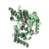
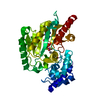
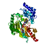
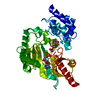
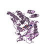
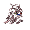
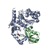
| Entry | Database: PDB / ID: 1bg0 | ||||||
|---|---|---|---|---|---|---|---|
| Title | TRANSITION STATE STRUCTURE OF ARGININE KINASE | ||||||
 Components Components | ARGININE KINASE | ||||||
 Keywords Keywords | KINASE / ARGININE KINASE / CREATINE KINASE / PHOSPHAGEN KINASE / TRANSITION STATE ANALOG / ADENOSINE TRIPHOSPHATE / TRANSFERASE | ||||||
| Function / homology |  Function and homology information Function and homology informationarginine kinase / arginine kinase activity / phosphocreatine biosynthetic process / creatine kinase activity / phosphorylation / : / ATP binding / cytoplasm Similarity search - Function | ||||||
| Biological species |  Limulus polyphemus (Atlantic horseshoe crab) Limulus polyphemus (Atlantic horseshoe crab) | ||||||
| Method |  X-RAY DIFFRACTION / molecular replacement, MIR / Resolution: 1.86 Å X-RAY DIFFRACTION / molecular replacement, MIR / Resolution: 1.86 Å | ||||||
 Authors Authors | Zhou, G. / Somasundaram, T. / Blanc, E. / Parthasarathy, G. / Ellington, W.R. / Chapman, M.S. | ||||||
 Citation Citation | Journal: Proc.Natl.Acad.Sci.USA / Year: 1998 Title: Transition state structure of arginine kinase: implications for catalysis of bimolecular reactions. Authors: Zhou, G. / Somasundaram, T. / Blanc, E. / Parthasarathy, G. / Ellington, W.R. / Chapman, M.S. #1: Journal: To be PublishedTitle: Critical Initial Real Space Refinement in Structure Determination of Arginine Kinase Authors: Zhou, G. / Somasundaram, T. / Blanc, E. / Chen, Z. / Chapman, M.S. #2: Journal: Protein Sci. / Year: 1997Title: Expression, Purification from Inclusion Bodies, and Crystal Characterization of a Transition State Analog Complex of Arginine Kinase: A Model for Studying Phosphagen Kinases Authors: Zhou, G. / Parthasarathy, G. / Somasundaram, T. / Ables, A. / Roy, L. / Strong, S.J. / Ellington, W.R. / Chapman, M.S. | ||||||
| History |
|
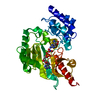
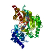
- Structure visualization
| Structure viewer | Molecule: MolmilJmol/JSmol |
|---|
- Downloads & links
-Download
| PDBx/mmCIF format | 1bg0.cif.gz | 92.6 KB | Display | PDBx/mmCIF format |
|---|---|---|---|---|
| PDB format | pdb1bg0.ent.gz | 68.4 KB | Display | PDB format |
| PDBx/mmJSON format | 1bg0.json.gz | Tree view | PDBx/mmJSON format | |
| Others |  Other downloads Other downloads |
-Validation report
| Arichive directory | https://data.pdbj.org/pub/pdb/validation_reports/bg/1bg0ftp://data.pdbj.org/pub/pdb/validation_reports/bg/1bg0 | HTTPS FTP |
|---|
-Related structure data
| Related structure data | 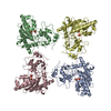 1crkS S: Starting model for refinement |
|---|---|
| Similar structure data |
-Links
 PDBj
PDBj
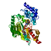
- Assembly
| Deposited unit | 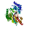
| ||||||||
|---|---|---|---|---|---|---|---|---|---|
| 1 |
| ||||||||
| Unit cell |
|
-Components
-Protein , 1 types, 1 molecules A
| #1: Protein | Mass: 40118.562 Da / Num. of mol.: 1 Source method: isolated from a genetically manipulated source Source: (gene. exp.) Limulus polyphemus (Atlantic horseshoe crab)Tissue: MUSCLE / Cellular location: CYTOPLASM / Gene: AK17 / Plasmid: PET-22B / Gene (production host): AK17 / Production host:  |
|---|
-Non-polymers , 5 types, 303 molecules 


| #2: Chemical |  Mass: 62.005 Da / Num. of mol.: 2 / Source method: obtained synthetically / Formula: NO3 Mass: 62.005 Da / Num. of mol.: 2 / Source method: obtained synthetically / Formula: NO3#3: Chemical | ChemComp-MG / |  Mass: 24.305 Da / Num. of mol.: 1 / Source method: obtained synthetically / Formula: Mg Mass: 24.305 Da / Num. of mol.: 1 / Source method: obtained synthetically / Formula: Mg#4: Chemical | ChemComp-ADP / |  Mass: 427.201 Da / Num. of mol.: 1 / Source method: obtained synthetically / Formula: C10H15N5O10P2 / Comment: ADP, energy-carrying molecule*YM Mass: 427.201 Da / Num. of mol.: 1 / Source method: obtained synthetically / Formula: C10H15N5O10P2 / Comment: ADP, energy-carrying molecule*YM#5: Chemical | ChemComp-DAR / |  Type: D-peptide linking / Mass: 175.209 Da / Num. of mol.: 1 / Source method: obtained synthetically / Formula: C6H15N4O2 Type: D-peptide linking / Mass: 175.209 Da / Num. of mol.: 1 / Source method: obtained synthetically / Formula: C6H15N4O2#6: Water | ChemComp-HOH / | Mass: 18.015 Da / Num. of mol.: 298 / Source method: isolated from a natural source / Formula: H2O |
|---|
-Experimental details
-Experiment
| Experiment | Method: X-RAY DIFFRACTION / Number of used crystals: 1 |
|---|
- Sample preparation
| Crystal | Density Matthews: 2.24 Å3/Da / Density % sol: 45 % Description: THE LARGE DOMAIN OF SUBUNIT A WAS USED AS A PROBE MODEL |
|---|---|
| Crystal grow | pH: 7.5 Details: PROTEIN WAS CRYSTALLIZED FROM 18% PEG 6000, 53 MM MGCL2, 2MM MGADP-, 25 MM KNO3, 10 MM ARGININE 0.5 MM DTT, 2.5 MM SODIUM AZIDE, 25 MM HEPES, PH 7.5, WITH 20 MG/ML OF PROTEIN CONCENTRATION. |
| Crystal | *PLUS |
| Crystal grow | *PLUS Method: microdialysis / Details: Zhou, G., (1997) Protein Sci., 6, 444. |
| Components of the solutions | *PLUS Conc.: 12 % / Common name: PEG6000 / Details: can be replaced by PEG1000 |
-Data collection
| Diffraction | Mean temperature: 100 K |
|---|---|
| Diffraction source | Source: ROTATING ANODE / Type: RIGAKU RUH2R / Wavelength: 1.5418 |
| Detector | Type: RIGAKU RAXIS IIC / Detector: IMAGE PLATE / Date: Sep 13, 1996 / Details: GRAPHITE MONOCHROMATOR OPTICS |
| Radiation | Monochromator: GRAPHITE(002) / Monochromatic (M) / Laue (L): M / Scattering type: x-ray |
| Radiation wavelength | Wavelength: 1.5418 Å / Relative weight: 1 |
| Reflection | Resolution: 1.86→35 Å / Num. obs: 34737 / % possible obs: 98.9 % / Observed criterion σ(I): 1 / Redundancy: 6.7 % / Biso Wilson estimate: 16.8 Å2 / Rmerge(I) obs: 0.047 / Net I/σ(I): 15.9 |
| Reflection shell | Resolution: 1.86→1.94 Å / Rmerge(I) obs: 0.157 / % possible all: 98.5 |
| Reflection shell | *PLUS % possible obs: 98.5 % |
- Processing
| Software |
| ||||||||||||||||||||||||||||||||||||||||||||||||||||||||||||
|---|---|---|---|---|---|---|---|---|---|---|---|---|---|---|---|---|---|---|---|---|---|---|---|---|---|---|---|---|---|---|---|---|---|---|---|---|---|---|---|---|---|---|---|---|---|---|---|---|---|---|---|---|---|---|---|---|---|---|---|---|---|
| Refinement | Method to determine structure: molecular replacement, MIR Starting model: 1CRK, MITOCHONDRIAL CREATINE KINASE Resolution: 1.86→5 Å / Data cutoff high absF: 10000 / Data cutoff low absF: 0.01 / Isotropic thermal model: GAUSSIAN DISTRIBUTION / Cross valid method: THROUGHOUT / σ(F): 2 Details: INITIAL LOW RESOLUTION REFINEMENT USED BOTH LOCAL REAL SPACE METHODS (CHAPMAN, M.S. ACTA CRYST., A51: 69-80 (1995); CHAPMAN, M.S. & BLANC, E. ACTA CRYST., 553: 203-6 (1997)).
| ||||||||||||||||||||||||||||||||||||||||||||||||||||||||||||
| Displacement parameters | Biso mean: 16.6 Å2 | ||||||||||||||||||||||||||||||||||||||||||||||||||||||||||||
| Refine analyze |
| ||||||||||||||||||||||||||||||||||||||||||||||||||||||||||||
| Refinement step | Cycle: LAST / Resolution: 1.86→5 Å
| ||||||||||||||||||||||||||||||||||||||||||||||||||||||||||||
| Refine LS restraints |
| ||||||||||||||||||||||||||||||||||||||||||||||||||||||||||||
| LS refinement shell | Resolution: 1.86→1.94 Å / Total num. of bins used: 8
| ||||||||||||||||||||||||||||||||||||||||||||||||||||||||||||
| Xplor file |
| ||||||||||||||||||||||||||||||||||||||||||||||||||||||||||||
| Software | *PLUS Name: X-PLOR / Version: 3.8 / Classification: refinement | ||||||||||||||||||||||||||||||||||||||||||||||||||||||||||||
| Refine LS restraints | *PLUS
|
