 Movie
Movie Controller


+ Open data

- Basic information
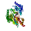
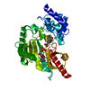
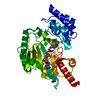
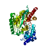
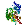
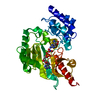
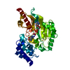
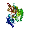
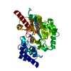
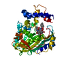
| Entry | Database: PDB / ID: 1p50 | ||||||
|---|---|---|---|---|---|---|---|
| Title | Transition state structure of an Arginine Kinase mutant | ||||||
 Components Components | Arginine kinase | ||||||
 Keywords Keywords | TRANSFERASE / phosphagen kinase / transition state | ||||||
| Function / homology |  Function and homology information Function and homology informationarginine kinase / arginine kinase activity / phosphocreatine biosynthetic process / creatine kinase activity / phosphorylation / : / ATP binding / cytoplasm Similarity search - Function | ||||||
| Biological species |  Limulus polyphemus (Atlantic horseshoe crab) Limulus polyphemus (Atlantic horseshoe crab) | ||||||
| Method |  X-RAY DIFFRACTION / MOLECULAR REPLACEMENT / Resolution: 2.8 Å X-RAY DIFFRACTION / MOLECULAR REPLACEMENT / Resolution: 2.8 Å | ||||||
 Authors Authors | Pruett, P.S. / Azzi, A. / Clark, S.A. / Yousef, M.S. / Gattis, J.L. / Somasundarum, T. / Ellington, W.R. / Chapman, M.S. | ||||||
 Citation Citation | Journal: J.Biol.Chem. / Year: 2003 Title: The putative catalytic bases have, at most, an accessory role in the mechanism of arginine kinase. Authors: Pruett, P.S. / Azzi, A. / Clark, S.A. / Yousef, M.S. / Gattis, J.L. / Somasundaram, T. / Ellington, W.R. / Chapman, M.S. #1: Journal: Proc.Natl.Acad.Sci.USA / Year: 1998Title: Transition state structure of arginine kinase: implications for catalysis of bimolecular reactions | ||||||
| History |
|
- Structure visualization
| Structure viewer | Molecule: MolmilJmol/JSmol |
|---|
- Downloads & links
-Download
| PDBx/mmCIF format | 1p50.cif.gz | 87.1 KB | Display | PDBx/mmCIF format |
|---|---|---|---|---|
| PDB format | pdb1p50.ent.gz | 64 KB | Display | PDB format |
| PDBx/mmJSON format | 1p50.json.gz | Tree view | PDBx/mmJSON format | |
| Others |  Other downloads Other downloads |
-Validation report
| Arichive directory | https://data.pdbj.org/pub/pdb/validation_reports/p5/1p50ftp://data.pdbj.org/pub/pdb/validation_reports/p5/1p50 | HTTPS FTP |
|---|
-Related structure data
| Related structure data |  1p52C 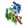 1bg0S S: Starting model for refinement C: citing same article ( |
|---|---|
| Similar structure data | |
| Other databases |
|
-Links
 PDBj
PDBj

- Assembly
| Deposited unit | 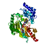
| ||||||||
|---|---|---|---|---|---|---|---|---|---|
| 1 |
| ||||||||
| Unit cell |
|
-Components
-Protein , 1 types, 1 molecules A
| #1: Protein | Mass: 40117.578 Da / Num. of mol.: 1 / Mutation: E103Q, G116A, D112G, E225Q Source method: isolated from a genetically manipulated source Source: (gene. exp.) Limulus polyphemus (Atlantic horseshoe crab)Plasmid: pET22b / Species (production host): Escherichia coli / Production host:  |
|---|
-Non-polymers , 5 types, 130 molecules 


| #2: Chemical | ChemComp-MG /  Mass: 24.305 Da / Num. of mol.: 1 / Source method: obtained synthetically / Formula: Mg Mass: 24.305 Da / Num. of mol.: 1 / Source method: obtained synthetically / Formula: Mg |
|---|---|
| #3: Chemical | ChemComp-ADP /  Mass: 427.201 Da / Num. of mol.: 1 / Source method: obtained synthetically / Formula: C10H15N5O10P2 / Comment: ADP, energy-carrying molecule*YM Mass: 427.201 Da / Num. of mol.: 1 / Source method: obtained synthetically / Formula: C10H15N5O10P2 / Comment: ADP, energy-carrying molecule*YM |
| #4: Chemical | ChemComp-NO3 /  Mass: 62.005 Da / Num. of mol.: 1 / Source method: obtained synthetically / Formula: NO3 Mass: 62.005 Da / Num. of mol.: 1 / Source method: obtained synthetically / Formula: NO3 |
| #5: Chemical | ChemComp-ARG /  Type: L-peptide linking / Mass: 175.209 Da / Num. of mol.: 1 / Source method: obtained synthetically / Formula: C6H15N4O2 Type: L-peptide linking / Mass: 175.209 Da / Num. of mol.: 1 / Source method: obtained synthetically / Formula: C6H15N4O2 |
| #6: Water | ChemComp-HOH / Mass: 18.015 Da / Num. of mol.: 126 / Source method: isolated from a natural source / Formula: H2O |
-Experimental details
-Experiment
| Experiment | Method: X-RAY DIFFRACTION / Number of used crystals: 4 |
|---|
- Sample preparation
| Crystal | Density Matthews: 2.24 Å3/Da / Density % sol: 44.54 % | |||||||||||||||||||||||||||||||||||||||||||||||||
|---|---|---|---|---|---|---|---|---|---|---|---|---|---|---|---|---|---|---|---|---|---|---|---|---|---|---|---|---|---|---|---|---|---|---|---|---|---|---|---|---|---|---|---|---|---|---|---|---|---|---|
| Crystal grow | Temperature: 277 K / Method: vapor diffusion, hanging drop / pH: 7.5 Details: PEG 6000, Hepes, magnesium chloride, ADP, nitrate, arginine, pH 7.5, VAPOR DIFFUSION, HANGING DROP, temperature 277K | |||||||||||||||||||||||||||||||||||||||||||||||||
| Crystal grow | *PLUS Temperature: 4 ℃ / Method: vapor diffusion | |||||||||||||||||||||||||||||||||||||||||||||||||
| Components of the solutions | *PLUS
|
-Data collection
| Diffraction |
| |||||||||||||||
|---|---|---|---|---|---|---|---|---|---|---|---|---|---|---|---|---|
| Diffraction source | Source: ROTATING ANODE / Type: RIGAKU / Wavelength: 1.5418 Å | |||||||||||||||
| Detector | Type: RIGAKU RAXIS / Detector: IMAGE PLATE / Date: Mar 10, 2000 / Details: mirrors | |||||||||||||||
| Radiation | Monochromator: rotating anode / Protocol: SINGLE WAVELENGTH / Monochromatic (M) / Laue (L): M / Scattering type: x-ray | |||||||||||||||
| Radiation wavelength | Wavelength: 1.5418 Å / Relative weight: 1 | |||||||||||||||
| Reflection | Resolution: 2.8→10 Å / Num. all: 9424 / Num. obs: 8914 / % possible obs: 94.6 % / Observed criterion σ(F): 2 / Observed criterion σ(I): 2 / Redundancy: 5.2 % / Rmerge(I) obs: 0.128 | |||||||||||||||
| Reflection shell | Resolution: 2.8→2.9 Å / % possible all: 92 | |||||||||||||||
| Reflection | *PLUS % possible obs: 97.4 % |
- Processing
| Software |
| |||||||||||||||||||||||||
|---|---|---|---|---|---|---|---|---|---|---|---|---|---|---|---|---|---|---|---|---|---|---|---|---|---|---|
| Refinement | Method to determine structure: MOLECULAR REPLACEMENT Starting model: PDB entry 1BG0 Resolution: 2.8→10 Å / Cross valid method: THROUGHOUT / σ(F): 2 / σ(I): 2 / Stereochemistry target values: Engh & Huber
| |||||||||||||||||||||||||
| Refine analyze | Luzzati sigma a free: 0.38 Å | |||||||||||||||||||||||||
| Refinement step | Cycle: LAST / Resolution: 2.8→10 Å
| |||||||||||||||||||||||||
| Refinement | *PLUS % reflection Rfree: 3-5 | |||||||||||||||||||||||||
| Solvent computation | *PLUS | |||||||||||||||||||||||||
| Displacement parameters | *PLUS |
