 Movie
Movie Controller

[English] 日本語
 Yorodumi
- PDB-6fes: Crystal structure of novel repeat protein BRIC2 fused to DARPin D12 -
+ Open data

- Basic information
| Entry | Database: PDB / ID: 6fes | ||||||
|---|---|---|---|---|---|---|---|
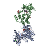
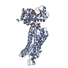
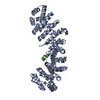
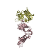
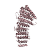
| Title | Crystal structure of novel repeat protein BRIC2 fused to DARPin D12 | ||||||
 Components Components | D12_BRIC2, a synthetic protein,D12_BRIC2, a synthetic protein | ||||||
 Keywords Keywords | DE NOVO PROTEIN / Novel fold / computational design / corrugated repeat | ||||||
| Function / homology | Ankyrin repeat-containing domain / Serine Threonine Protein Phosphatase 5, Tetratricopeptide repeat / Alpha Horseshoe / Mainly Alpha Function and homology information Function and homology information | ||||||
| Biological species | synthetic construct (others) | ||||||
| Method |  X-RAY DIFFRACTION / SYNCHROTRON / MOLECULAR REPLACEMENT / Resolution: 3 Å X-RAY DIFFRACTION / SYNCHROTRON / MOLECULAR REPLACEMENT / Resolution: 3 Å | ||||||
 Authors Authors | ElGamacy, M. / Coles, M. / Ernst, P. / Zhu, H. / Hartmann, M.D. / Plueckthun, A. / Lupas, A. | ||||||
 Citation Citation | Journal: ACS Synth Biol / Year: 2018 Title: An Interface-Driven Design Strategy Yields a Novel, Corrugated Protein Architecture. Authors: ElGamacy, M. / Coles, M. / Ernst, P. / Zhu, H. / Hartmann, M.D. / Pluckthun, A. / Lupas, A.N. | ||||||
| History |
|
- Structure visualization
| Structure viewer | Molecule: MolmilJmol/JSmol |
|---|
- Downloads & links
-Download
| PDBx/mmCIF format | 6fes.cif.gz | 819.7 KB | Display | PDBx/mmCIF format |
|---|---|---|---|---|
| PDB format | pdb6fes.ent.gz | 704.6 KB | Display | PDB format |
| PDBx/mmJSON format | 6fes.json.gz | Tree view | PDBx/mmJSON format | |
| Others |  Other downloads Other downloads |
-Validation report
| Arichive directory | https://data.pdbj.org/pub/pdb/validation_reports/fe/6fesftp://data.pdbj.org/pub/pdb/validation_reports/fe/6fes | HTTPS FTP |
|---|
-Related structure data
| Related structure data |  6ff6C 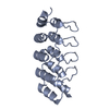 4ydwS S: Starting model for refinement C: citing same article ( |
|---|---|
| Similar structure data |


-Links
 PDBj
PDBj
- Assembly
| Deposited unit | 
| ||||||||
|---|---|---|---|---|---|---|---|---|---|
| 1 | 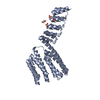
| ||||||||
| 2 | 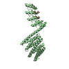
| ||||||||
| 3 | 
| ||||||||
| 4 | 
| ||||||||
| Unit cell |
|
-Components
| #1: Protein | Mass: 41061.582 Da / Num. of mol.: 4 Fragment: D12 residues 11-157, linker residues 158-172,Residues 173-375,D12 residues 11-157, linker residues 158-172,Residues 173-375 Source method: isolated from a genetically manipulated source Source: (gene. exp.) synthetic construct (others) / Production host:  #2: Chemical | ChemComp-SO4 /   Mass: 96.063 Da / Num. of mol.: 9 / Source method: obtained synthetically / Formula: SO4 Mass: 96.063 Da / Num. of mol.: 9 / Source method: obtained synthetically / Formula: SO4#3: Chemical | ChemComp-EDO /   Mass: 62.068 Da / Num. of mol.: 7 / Source method: obtained synthetically / Formula: C2H6O2 Mass: 62.068 Da / Num. of mol.: 7 / Source method: obtained synthetically / Formula: C2H6O2#4: Water | ChemComp-HOH / |  Mass: 18.015 Da / Num. of mol.: 51 / Source method: isolated from a natural source / Formula: H2O Mass: 18.015 Da / Num. of mol.: 51 / Source method: isolated from a natural source / Formula: H2OHas protein modification | N | |
|---|
-Experimental details
-Experiment
| Experiment | Method: X-RAY DIFFRACTION / Number of used crystals: 1 |
|---|
- Sample preparation
| Crystal | Density Matthews: 2.5 Å3/Da / Density % sol: 50.85 % |
|---|---|
| Crystal grow | Temperature: 277.15 K / Method: vapor diffusion Details: 0.2 M Ammonium sulfate, 0.1M Bis-Tris pH 5.5, 25 % (w/v) PEG 3350 |
-Data collection
| Diffraction | Mean temperature: 100 K |
|---|---|
| Diffraction source | Source: SYNCHROTRON / Site: SLS  / Beamline: X06SA / Wavelength: 1 Å / Beamline: X06SA / Wavelength: 1 Å |
| Detector | Type: DECTRIS EIGER X 16M / Detector: PIXEL / Date: Dec 4, 2017 |
| Radiation | Protocol: SINGLE WAVELENGTH / Monochromatic (M) / Laue (L): M / Scattering type: x-ray |
| Radiation wavelength | Wavelength: 1 Å / Relative weight: 1 |
| Reflection | Resolution: 3→49.33 Å / Num. obs: 31567 / % possible obs: 99 % / Redundancy: 5.6 % / Rmerge(I) obs: 0.177 / Net I/σ(I): 7.6 |
| Reflection shell | Resolution: 3→3.08 Å / Rmerge(I) obs: 1.308 / Mean I/σ(I) obs: 2.15 / % possible all: 99.8 |
- Processing
| Software |
| ||||||||||||||||||||||||||||||||||||||||||||||||||||||||||||||||||||||||||||||||||||
|---|---|---|---|---|---|---|---|---|---|---|---|---|---|---|---|---|---|---|---|---|---|---|---|---|---|---|---|---|---|---|---|---|---|---|---|---|---|---|---|---|---|---|---|---|---|---|---|---|---|---|---|---|---|---|---|---|---|---|---|---|---|---|---|---|---|---|---|---|---|---|---|---|---|---|---|---|---|---|---|---|---|---|---|---|---|
| Refinement | Method to determine structure: MOLECULAR REPLACEMENT Starting model: one DARPin chain from structure 4ydw Resolution: 3→44.895 Å / SU ML: 0.52 / Cross valid method: FREE R-VALUE / σ(F): 1.97 / Phase error: 34.35 / Stereochemistry target values: ML
| ||||||||||||||||||||||||||||||||||||||||||||||||||||||||||||||||||||||||||||||||||||
| Solvent computation | Shrinkage radii: 0.9 Å / VDW probe radii: 1.11 Å / Solvent model: FLAT BULK SOLVENT MODEL | ||||||||||||||||||||||||||||||||||||||||||||||||||||||||||||||||||||||||||||||||||||
| Refinement step | Cycle: LAST / Resolution: 3→44.895 Å
| ||||||||||||||||||||||||||||||||||||||||||||||||||||||||||||||||||||||||||||||||||||
| Refine LS restraints |
| ||||||||||||||||||||||||||||||||||||||||||||||||||||||||||||||||||||||||||||||||||||
| LS refinement shell |
| ||||||||||||||||||||||||||||||||||||||||||||||||||||||||||||||||||||||||||||||||||||
| Refinement TLS params. | Method: refined / Origin x: -13.3111 Å / Origin y: 2.9493 Å / Origin z: 19.6649 Å
| ||||||||||||||||||||||||||||||||||||||||||||||||||||||||||||||||||||||||||||||||||||
| Refinement TLS group | Selection details: all |
