 Movie
Movie Controller


+ Open data

- Basic information
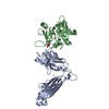
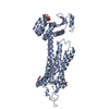
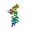
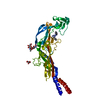
| Entry | Database: PDB / ID: 1l0x | ||||||
|---|---|---|---|---|---|---|---|
| Title | TCR beta chain complexed with streptococcal superantigen SpeA | ||||||
 Components Components |
| ||||||
 Keywords Keywords | IMMUNE SYSTEM / TCR / superantigen | ||||||
| Function / homology |  Function and homology information Function and homology informationalpha-beta T cell receptor complex / toxin activity / extracellular region Similarity search - Function | ||||||
| Biological species |   Streptococcus pyogenes (bacteria) Streptococcus pyogenes (bacteria) | ||||||
| Method |  X-RAY DIFFRACTION / SYNCHROTRON / MOLECULAR REPLACEMENT / Resolution: 2.8 Å X-RAY DIFFRACTION / SYNCHROTRON / MOLECULAR REPLACEMENT / Resolution: 2.8 Å | ||||||
 Authors Authors | Li, H. / Sundberg, E.J. / Mariuzza, R.A. | ||||||
 Citation Citation | Journal: Structure / Year: 2002 Title: Structures of two streptococcal superantigens bound to TCR beta chains reveal diversity in the architecture of T cell signaling complexes. Authors: Sundberg, E.J. / Li, H. / Llera, A.S. / McCormick, J.K. / Tormo, J. / Schlievert, P.M. / Karjalainen, K. / Mariuzza, R.A. | ||||||
| History |
| ||||||
| Remark 999 | SEQUENCE Chains A and C are not D10 TCR, genbank entry AAB41230. They are rather 14.3.d TCR, whose ...SEQUENCE Chains A and C are not D10 TCR, genbank entry AAB41230. They are rather 14.3.d TCR, whose sequence has not been deposited. The two are slightly different and hence give rise to the four SEQADV. |
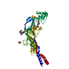
- Structure visualization
| Structure viewer | Molecule: MolmilJmol/JSmol |
|---|
- Downloads & links
-Download
| PDBx/mmCIF format | 1l0x.cif.gz | 192.7 KB | Display | PDBx/mmCIF format |
|---|---|---|---|---|
| PDB format | pdb1l0x.ent.gz | 151.9 KB | Display | PDB format |
| PDBx/mmJSON format | 1l0x.json.gz | Tree view | PDBx/mmJSON format | |
| Others |  Other downloads Other downloads |
-Validation report
| Arichive directory | https://data.pdbj.org/pub/pdb/validation_reports/l0/1l0xftp://data.pdbj.org/pub/pdb/validation_reports/l0/1l0x | HTTPS FTP |
|---|
-Related structure data
| Related structure data | 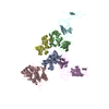 1ktkC  1l0yC 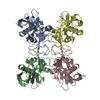 1b1zS 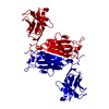 1becS C: citing same article ( S: Starting model for refinement |
|---|---|
| Similar structure data |


-Links
 PDBj
PDBj
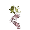
- Assembly
| Deposited unit | 
| ||||||||
|---|---|---|---|---|---|---|---|---|---|
| 1 | 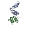
| ||||||||
| 2 | 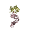
| ||||||||
| Unit cell |
|
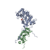
-Components
| #1: Protein | Mass: 26568.490 Da / Num. of mol.: 2 / Mutation: N24Q, N74Q, N121Q Source method: isolated from a genetically manipulated source Source: (gene. exp.) #2: Protein | Mass: 25803.932 Da / Num. of mol.: 2 / Mutation: C90S Source method: isolated from a genetically manipulated source Source: (gene. exp.) Streptococcus pyogenes (bacteria) / Production host: #3: Chemical |   Mass: 92.094 Da / Num. of mol.: 3 / Source method: obtained synthetically / Formula: C3H8O3 Mass: 92.094 Da / Num. of mol.: 3 / Source method: obtained synthetically / Formula: C3H8O3#4: Water | ChemComp-HOH / |  Mass: 18.015 Da / Num. of mol.: 181 / Source method: isolated from a natural source / Formula: H2O Mass: 18.015 Da / Num. of mol.: 181 / Source method: isolated from a natural source / Formula: H2OHas protein modification | Y | |
|---|
-Experimental details
-Experiment
| Experiment | Method: X-RAY DIFFRACTION / Number of used crystals: 1 |
|---|
- Sample preparation
| Crystal | Density Matthews: 2.65 Å3/Da / Density % sol: 53.64 % | |||||||||||||||||||||||||||||||||||
|---|---|---|---|---|---|---|---|---|---|---|---|---|---|---|---|---|---|---|---|---|---|---|---|---|---|---|---|---|---|---|---|---|---|---|---|---|
| Crystal grow | Temperature: 298 K / Method: vapor diffusion, hanging drop / pH: 8 Details: 15% PEG8000, 0.2M MgCl2, 0.1M Tris, pH 8.0, VAPOR DIFFUSION, HANGING DROP, temperature 298K | |||||||||||||||||||||||||||||||||||
| Crystal grow | *PLUS | |||||||||||||||||||||||||||||||||||
| Components of the solutions | *PLUS
|
-Data collection
| Diffraction | Mean temperature: 100 K |
|---|---|
| Diffraction source | Source: SYNCHROTRON / Site: APS  / Beamline: 19-ID / Wavelength: 0.978 Å / Beamline: 19-ID / Wavelength: 0.978 Å |
| Detector | Type: ADSC QUANTUM 4 / Detector: CCD / Date: Feb 20, 2000 |
| Radiation | Monochromator: GRAPHITE / Protocol: SINGLE WAVELENGTH / Monochromatic (M) / Laue (L): M / Scattering type: x-ray |
| Radiation wavelength | Wavelength: 0.978 Å / Relative weight: 1 |
| Reflection | Resolution: 2.8→40.78 Å / Num. all: 38028 / Num. obs: 26926 / % possible obs: 99.1 % / Observed criterion σ(F): 1 / Observed criterion σ(I): 1 / Redundancy: 2.99 % / Biso Wilson estimate: 61.66 Å2 / Rmerge(I) obs: 0.121 / Rsym value: 0.121 / Net I/σ(I): 9.6 |
| Reflection shell | Resolution: 2.8→2.9 Å / Rmerge(I) obs: 0.373 / Mean I/σ(I) obs: 3.6 / Num. unique obs: 2696 / Rsym value: 0.373 / % possible all: 99.8 |
| Reflection | *PLUS % possible obs: 99.1 % / Num. measured all: 80520 |
| Reflection shell | *PLUS % possible obs: 99.8 % |
- Processing
| Software |
| |||||||||||||||||||||||||
|---|---|---|---|---|---|---|---|---|---|---|---|---|---|---|---|---|---|---|---|---|---|---|---|---|---|---|
| Refinement | Method to determine structure: MOLECULAR REPLACEMENT Starting model: PDB entry 1BEC and 1B1Z Resolution: 2.8→40.78 Å / Isotropic thermal model: Isotropic / Cross valid method: THROUGHOUT / σ(F): 0 / σ(I): 0 / Stereochemistry target values: Engh & Huber
| |||||||||||||||||||||||||
| Refinement step | Cycle: LAST / Resolution: 2.8→40.78 Å
| |||||||||||||||||||||||||
| Refine LS restraints |
| |||||||||||||||||||||||||
| Refinement | *PLUS % reflection Rfree: 4.9 % / Rfactor obs: 0.232 | |||||||||||||||||||||||||
| Solvent computation | *PLUS | |||||||||||||||||||||||||
| Displacement parameters | *PLUS | |||||||||||||||||||||||||
| LS refinement shell | *PLUS Highest resolution: 2.8 Å / Lowest resolution: 2.9 Å / Rfactor Rfree: 0.459 / Rfactor Rwork: 0.397 / Rfactor obs: 0.397 |
