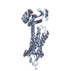
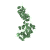
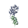
Entry Database : PDB / ID : 4rt7Title Crystal Structure of FLT3 with a small molecule inhibitor Receptor-type tyrosine-protein kinase FLT3 Keywords / / / / / Function / homology Function Domain/homology Component
/ / / / / / / / / / / / / / / / / / / / / / / / / / / / / / / / / / / / / / / / / / / / / / / / / / / / / / / / / / / / / / / / / / / / / / / / / / / / / / / / / / / / / / / / / / / / / / / / / / / / / / / / / / / / Biological species Homo sapiens (human)Method / / / Resolution : 3.1 Å Authors Zhang, Y. / Zhang, C. Journal : Cancer Discov / Year : 2015Title : Characterizing and Overriding the Structural Mechanism of the Quizartinib-Resistant FLT3 "Gatekeeper" F691L Mutation with PLX3397.Authors: Smith, C.C. / Zhang, C. / Lin, K.C. / Lasater, E.A. / Zhang, Y. / Massi, E. / Damon, L.E. / Pendleton, M. / Bashir, A. / Sebra, R. / Perl, A. / Kasarskis, A. / Shellooe, R. / Tsang, G. / ... Authors : Smith, C.C. / Zhang, C. / Lin, K.C. / Lasater, E.A. / Zhang, Y. / Massi, E. / Damon, L.E. / Pendleton, M. / Bashir, A. / Sebra, R. / Perl, A. / Kasarskis, A. / Shellooe, R. / Tsang, G. / Carias, H. / Powell, B. / Burton, E.A. / Matusow, B. / Zhang, J. / Spevak, W. / Ibrahim, P.N. / Le, M.H. / Hsu, H.H. / Habets, G. / West, B.L. / Bollag, G. / Shah, N.P. History Deposition Nov 13, 2014 Deposition site / Processing site Revision 1.0 Apr 22, 2015 Provider / Type Revision 1.1 Jun 17, 2015 Group Revision 1.2 Nov 6, 2024 Group Data collection / Database references ... Data collection / Database references / Derived calculations / Structure summary Category chem_comp / chem_comp_atom ... chem_comp / chem_comp_atom / chem_comp_bond / database_2 / pdbx_entry_details / pdbx_modification_feature / struct_ref_seq_dif / struct_site Item _chem_comp.pdbx_synonyms / _database_2.pdbx_DOI ... _chem_comp.pdbx_synonyms / _database_2.pdbx_DOI / _database_2.pdbx_database_accession / _struct_ref_seq_dif.details / _struct_site.pdbx_auth_asym_id / _struct_site.pdbx_auth_comp_id / _struct_site.pdbx_auth_seq_id
Show all Show less
 Movie
Movie Controller


 Open data
 Basic information
 Components
 Keywords
 Function and homology information
 Homo sapiens (human)
 X-RAY DIFFRACTION /
  Authors
 Citation
 Structure visualization
 Downloads & links
 Other downloads
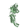


 PDBj
PDBj


 Assembly


 Spodoptera frugiperda (fall armyworm)
 Mass: 560.667 Da / Num. of mol.: 1 / Source method: obtained synthetically / Formula: C29H32N6O4S
 Mass: 18.015 Da / Num. of mol.: 1 / Source method: isolated from a natural source / Formula: H2O
 Sample preparation
 / Beamline: 8.3.1 / Wavelength: 1.1159 Å
 Processing