 Movie
Movie Controller

[English] 日本語
 Yorodumi
- PDB-5u1x: Crystal structure of the ATP-gated P2X7 ion channel bound to allo... -
+ Open data

- Basic information
| Entry | Database: PDB / ID: 5u1x | |||||||||
|---|---|---|---|---|---|---|---|---|---|---|
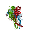
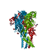
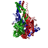
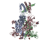
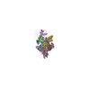
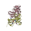
| Title | Crystal structure of the ATP-gated P2X7 ion channel bound to allosteric antagonist JNJ47965567 | |||||||||
 Components Components | P2X purinoceptor | |||||||||
 Keywords Keywords | MEMBRANE PROTEIN / membrane protein: ATP-gated ion channel: allosteric antagonist bound: closed state | |||||||||
| Function / homology |  Function and homology information Function and homology informationNAD transport / gamma-aminobutyric acid secretion / phagolysosome assembly / phospholipid transfer to membrane / plasma membrane organization / extracellularly ATP-gated monoatomic cation channel activity / positive regulation of interleukin-1 alpha production / purinergic nucleotide receptor activity / collagen metabolic process / positive regulation of prostaglandin secretion ...NAD transport / gamma-aminobutyric acid secretion / phagolysosome assembly / phospholipid transfer to membrane / plasma membrane organization / extracellularly ATP-gated monoatomic cation channel activity / positive regulation of interleukin-1 alpha production / purinergic nucleotide receptor activity / collagen metabolic process / positive regulation of prostaglandin secretion / pore complex assembly / negative regulation of cell volume / T cell apoptotic process / positive regulation of gamma-aminobutyric acid secretion / bleb assembly / mitochondrial depolarization / vesicle budding from membrane / positive regulation of T cell apoptotic process / prostaglandin secretion / response to fluid shear stress / glutamate secretion / cellular response to dsRNA / ceramide biosynthetic process / negative regulation of bone resorption / skeletal system morphogenesis / positive regulation of macrophage cytokine production / phospholipid translocation / response to zinc ion / positive regulation of glutamate secretion / response to ATP / T cell homeostasis / positive regulation of mitochondrial depolarization / membrane protein ectodomain proteolysis / positive regulation of NLRP3 inflammasome complex assembly / synaptic vesicle exocytosis / protein secretion / response to electrical stimulus / T cell proliferation / positive regulation of bone mineralization / response to mechanical stimulus / extrinsic apoptotic signaling pathway / negative regulation of MAPK cascade / release of sequestered calcium ion into cytosol / reactive oxygen species metabolic process / homeostasis of number of cells within a tissue / sensory perception of pain / positive regulation of interleukin-1 beta production / T cell mediated cytotoxicity / protein catabolic process / positive regulation of protein secretion / neuromuscular junction / mitochondrion organization / lipopolysaccharide binding / protein processing / positive regulation of T cell mediated cytotoxicity / response to calcium ion / positive regulation of interleukin-6 production / cell morphogenesis / cell-cell junction / MAPK cascade / presynapse / response to lipopolysaccharide / positive regulation of MAPK cascade / postsynapse / defense response to Gram-positive bacterium / response to xenobiotic stimulus / inflammatory response / external side of plasma membrane / neuronal cell body / mitochondrion / ATP binding / identical protein binding Similarity search - Function | |||||||||
| Biological species |  | |||||||||
| Method |  X-RAY DIFFRACTION / SYNCHROTRON / MOLECULAR REPLACEMENT / Resolution: 3.201 Å X-RAY DIFFRACTION / SYNCHROTRON / MOLECULAR REPLACEMENT / Resolution: 3.201 Å | |||||||||
 Authors Authors | Karasawa, A. / Kawate, T. | |||||||||
| Funding support |  United States, 2items United States, 2items
| |||||||||
 Citation Citation | Journal: Elife / Year: 2016 Title: Structural basis for subtype-specific inhibition of the P2X7 receptor. Authors: Karasawa, A. / Kawate, T. | |||||||||
| History |
|
- Structure visualization
| Structure viewer | Molecule: MolmilJmol/JSmol |
|---|
- Downloads & links
-Download
| PDBx/mmCIF format | 5u1x.cif.gz | 81.9 KB | Display | PDBx/mmCIF format |
|---|---|---|---|---|
| PDB format | pdb5u1x.ent.gz | 57.7 KB | Display | PDB format |
| PDBx/mmJSON format | 5u1x.json.gz | Tree view | PDBx/mmJSON format | |
| Others |  Other downloads Other downloads |
-Validation report
| Arichive directory | https://data.pdbj.org/pub/pdb/validation_reports/u1/5u1xftp://data.pdbj.org/pub/pdb/validation_reports/u1/5u1x | HTTPS FTP |
|---|
-Related structure data
| Related structure data |  5u1lC  5u1uC  5u1vC  5u1wC 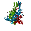 5u1yC  5u2hC C: citing same article ( |
|---|---|
| Similar structure data |

-Links
 PDBj
PDBj- Assembly
| Deposited unit | 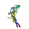
| ||||||||
|---|---|---|---|---|---|---|---|---|---|
| 1 | 
| ||||||||
| Unit cell |
|
-Components
| #1: Protein | Mass: 38716.234 Da / Num. of mol.: 1 Source method: isolated from a genetically manipulated source Details: Both the N- and C-termini are disordered and the electron density was not well-defined. Source: (gene. exp.)   Spodoptera frugiperda (fall armyworm) / References: UniProt: G1M6C4 Spodoptera frugiperda (fall armyworm) / References: UniProt: G1M6C4 | ||||
|---|---|---|---|---|---|
| #2: Polysaccharide | 2-acetamido-2-deoxy-beta-D-glucopyranose-(1-4)-2-acetamido-2-deoxy-beta-D-glucopyranose Source method: isolated from a genetically manipulated source | ||||
| #3: Sugar |   Type: D-saccharide, beta linking / Mass: 221.208 Da / Num. of mol.: 2 Type: D-saccharide, beta linking / Mass: 221.208 Da / Num. of mol.: 2Source method: isolated from a genetically manipulated source Formula: C8H15NO6 #4: Chemical | ChemComp-7RV / |   Mass: 488.644 Da / Num. of mol.: 1 / Source method: obtained synthetically / Formula: C28H32N4O2S Mass: 488.644 Da / Num. of mol.: 1 / Source method: obtained synthetically / Formula: C28H32N4O2SHas protein modification | Y | |
-Experimental details
-Experiment
| Experiment | Method: X-RAY DIFFRACTION / Number of used crystals: 1 |
|---|
- Sample preparation
| Crystal | Density Matthews: 5.22 Å3/Da / Density % sol: 76.42 % |
|---|---|
| Crystal grow | Temperature: 277 K / Method: vapor diffusion, hanging drop Details: 100 mM HEPES or Tris (pH 6.0-7.5), 100 mM NaCl, 4% ethylene glycol, 15% glycerol, 27-32% PEG-400 or 31-36% PEG-300, 0.1 mg/mL lipid mixture (60% POPE, 20% POPG, and 20% cholesterol), and 1 mM JNJ-47965567 PH range: 6.0-7.5 |
-Data collection
| Diffraction | Mean temperature: 100 K | ||||||||||||||||||||||||||||||
|---|---|---|---|---|---|---|---|---|---|---|---|---|---|---|---|---|---|---|---|---|---|---|---|---|---|---|---|---|---|---|---|
| Diffraction source | Source: SYNCHROTRON / Site: CHESS / Beamline: F1 / Wavelength: 0.9774 Å | ||||||||||||||||||||||||||||||
| Detector | Type: DECTRIS PILATUS3 S 6M / Detector: PIXEL / Date: Jul 10, 2015 | ||||||||||||||||||||||||||||||
| Radiation | Protocol: SINGLE WAVELENGTH / Monochromatic (M) / Laue (L): M / Scattering type: x-ray | ||||||||||||||||||||||||||||||
| Radiation wavelength | Wavelength: 0.9774 Å / Relative weight: 1 | ||||||||||||||||||||||||||||||
| Reflection | Resolution: 3.1→48.88 Å / Num. obs: 14815 / % possible obs: 100 % / Redundancy: 10.1 % / Biso Wilson estimate: 107.11 Å2 / CC1/2: 0.996 / Rmerge(I) obs: 0.134 / Net I/σ(I): 12.1 | ||||||||||||||||||||||||||||||
| Reflection shell | Diffraction-ID: 1 / Rejects: _
|
- Processing
| Software |
| ||||||||||||||||||||||||||||||||||||||||||||||||||||||||||||||||||
|---|---|---|---|---|---|---|---|---|---|---|---|---|---|---|---|---|---|---|---|---|---|---|---|---|---|---|---|---|---|---|---|---|---|---|---|---|---|---|---|---|---|---|---|---|---|---|---|---|---|---|---|---|---|---|---|---|---|---|---|---|---|---|---|---|---|---|---|
| Refinement | Method to determine structure: MOLECULAR REPLACEMENT Starting model: A740003 bound P2X7 Resolution: 3.201→48.88 Å / SU ML: 0.48 / Cross valid method: FREE R-VALUE / σ(F): 1.34 / Phase error: 27.66
| ||||||||||||||||||||||||||||||||||||||||||||||||||||||||||||||||||
| Solvent computation | Shrinkage radii: 0.9 Å / VDW probe radii: 1.11 Å | ||||||||||||||||||||||||||||||||||||||||||||||||||||||||||||||||||
| Displacement parameters | Biso max: 201.46 Å2 / Biso mean: 103.6362 Å2 / Biso min: 53.9 Å2 | ||||||||||||||||||||||||||||||||||||||||||||||||||||||||||||||||||
| Refinement step | Cycle: final / Resolution: 3.201→48.88 Å
| ||||||||||||||||||||||||||||||||||||||||||||||||||||||||||||||||||
| Refine LS restraints |
| ||||||||||||||||||||||||||||||||||||||||||||||||||||||||||||||||||
| LS refinement shell | Refine-ID: X-RAY DIFFRACTION / Total num. of bins used: 10 / % reflection obs: 100 %
|
