 Movie
Movie Controller

[English] 日本語
 Yorodumi
- PDB-6f4w: Crystal structure of H. pylori purine nucleoside phosphorylase in... -
+ Open data

- Basic information
| Entry | Database: PDB / ID: 6f4w | ||||||||||||
|---|---|---|---|---|---|---|---|---|---|---|---|---|---|
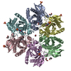
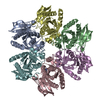
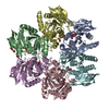
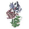
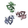
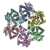
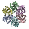
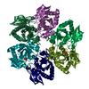
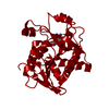
| Title | Crystal structure of H. pylori purine nucleoside phosphorylase in complex with formycin A | ||||||||||||
 Components Components | Purine nucleoside phosphorylase DeoD-type | ||||||||||||
 Keywords Keywords | HYDROLASE / Purine nucleoside phosphorylase / H. pylori / formycin A | ||||||||||||
| Function / homology |  Function and homology information Function and homology informationpurine-nucleoside phosphorylase activity / purine-nucleoside phosphorylase / purine nucleoside catabolic process / cytosol Similarity search - Function | ||||||||||||
| Biological species |   Helicobacter pylori (bacteria) Helicobacter pylori (bacteria) | ||||||||||||
| Method |  X-RAY DIFFRACTION / SYNCHROTRON / MOLECULAR REPLACEMENT / Resolution: 2.293 Å X-RAY DIFFRACTION / SYNCHROTRON / MOLECULAR REPLACEMENT / Resolution: 2.293 Å | ||||||||||||
 Authors Authors | Stefanic, Z. | ||||||||||||
| Funding support |  Croatia, Croatia,  Poland, 3items Poland, 3items
| ||||||||||||
 Citation Citation | Journal: FEBS J. / Year: 2018 Title: Helicobacter pylori purine nucleoside phosphorylase shows new distribution patterns of open and closed active site conformations and unusual biochemical features. Authors: Narczyk, M. / Bertosa, B. / Papa, L. / Vukovic, V. / Lescic Asler, I. / Wielgus-Kutrowska, B. / Bzowska, A. / Luic, M. / Stefanic, Z. | ||||||||||||
| History |
|
- Structure visualization
| Structure viewer | Molecule: MolmilJmol/JSmol |
|---|
- Downloads & links
-Download
| PDBx/mmCIF format | 6f4w.cif.gz | 284.8 KB | Display | PDBx/mmCIF format |
|---|---|---|---|---|
| PDB format | pdb6f4w.ent.gz | 230.4 KB | Display | PDB format |
| PDBx/mmJSON format | 6f4w.json.gz | Tree view | PDBx/mmJSON format | |
| Others |  Other downloads Other downloads |
-Validation report
| Arichive directory | https://data.pdbj.org/pub/pdb/validation_reports/f4/6f4wftp://data.pdbj.org/pub/pdb/validation_reports/f4/6f4w | HTTPS FTP |
|---|
-Related structure data
| Related structure data |  5lu0C  6f4xC  6f52C  6f5aC  6f5iC  5mx4S C: citing same article ( S: Starting model for refinement |
|---|---|
| Similar structure data |
-Links
 PDBj
PDBj- Assembly
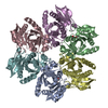
| Deposited unit | 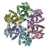
| ||||||||
|---|---|---|---|---|---|---|---|---|---|
| 1 |
| ||||||||
| Unit cell |
|
-Components
| #1: Protein | Mass: 25818.105 Da / Num. of mol.: 6 Source method: isolated from a genetically manipulated source Source: (gene. exp.) Helicobacter pylori (bacteria) / Strain: ATCC 700392 / 26695 / Gene: deoD, HP_1178 / Plasmid: pET21b / Production host: References: UniProt: P56463, purine-nucleoside phosphorylase #2: Chemical | ChemComp-FMC / (   Mass: 267.241 Da / Num. of mol.: 6 / Source method: obtained synthetically / Formula: C10H13N5O4 Mass: 267.241 Da / Num. of mol.: 6 / Source method: obtained synthetically / Formula: C10H13N5O4#3: Water | ChemComp-HOH / |  Mass: 18.015 Da / Num. of mol.: 422 / Source method: isolated from a natural source / Formula: H2O Mass: 18.015 Da / Num. of mol.: 422 / Source method: isolated from a natural source / Formula: H2O |
|---|
-Experimental details
-Experiment
| Experiment | Method: X-RAY DIFFRACTION / Number of used crystals: 1 |
|---|
- Sample preparation
| Crystal | Density Matthews: 2.37 Å3/Da / Density % sol: 48.16 % |
|---|---|
| Crystal grow | Temperature: 291 K / Method: vapor diffusion, hanging drop / pH: 7 / Details: 0.1M TRIS-HCl, 10% PEG 8000 |
-Data collection
| Diffraction | Mean temperature: 100 K | ||||||||||||||||||||||||||||||||||||||||||||||||||||||||||||||||||||||||||||||||||||||||
|---|---|---|---|---|---|---|---|---|---|---|---|---|---|---|---|---|---|---|---|---|---|---|---|---|---|---|---|---|---|---|---|---|---|---|---|---|---|---|---|---|---|---|---|---|---|---|---|---|---|---|---|---|---|---|---|---|---|---|---|---|---|---|---|---|---|---|---|---|---|---|---|---|---|---|---|---|---|---|---|---|---|---|---|---|---|---|---|---|---|
| Diffraction source | Source: SYNCHROTRON / Site: ELETTRA  / Beamline: 5.2R / Wavelength: 1 Å / Beamline: 5.2R / Wavelength: 1 Å | ||||||||||||||||||||||||||||||||||||||||||||||||||||||||||||||||||||||||||||||||||||||||
| Detector | Type: DECTRIS PILATUS3 2M / Detector: PIXEL / Date: Sep 5, 2016 | ||||||||||||||||||||||||||||||||||||||||||||||||||||||||||||||||||||||||||||||||||||||||
| Radiation | Protocol: SINGLE WAVELENGTH / Monochromatic (M) / Laue (L): M / Scattering type: x-ray | ||||||||||||||||||||||||||||||||||||||||||||||||||||||||||||||||||||||||||||||||||||||||
| Radiation wavelength | Wavelength: 1 Å / Relative weight: 1 | ||||||||||||||||||||||||||||||||||||||||||||||||||||||||||||||||||||||||||||||||||||||||
| Reflection | Resolution: 2.293→46.081 Å / Num. obs: 66682 / % possible obs: 99.4 % / Redundancy: 4.1 % / Biso Wilson estimate: 40.47 Å2 / Rpim(I) all: 0.083 / Rrim(I) all: 0.168 / Rsym value: 0.145 / Net I/av σ(I): 3.598 / Net I/σ(I): 5.8 | ||||||||||||||||||||||||||||||||||||||||||||||||||||||||||||||||||||||||||||||||||||||||
| Reflection shell | Diffraction-ID: 1
|
- Processing
| Software |
| |||||||||||||||||||||||||||||||||||||||||||||||||||||||||||||||||||||||||||||||||||||||||||||||||||||||||||||||||||||||||||||||||||||||||||||||||||||||||||||||||||||||||||||||
|---|---|---|---|---|---|---|---|---|---|---|---|---|---|---|---|---|---|---|---|---|---|---|---|---|---|---|---|---|---|---|---|---|---|---|---|---|---|---|---|---|---|---|---|---|---|---|---|---|---|---|---|---|---|---|---|---|---|---|---|---|---|---|---|---|---|---|---|---|---|---|---|---|---|---|---|---|---|---|---|---|---|---|---|---|---|---|---|---|---|---|---|---|---|---|---|---|---|---|---|---|---|---|---|---|---|---|---|---|---|---|---|---|---|---|---|---|---|---|---|---|---|---|---|---|---|---|---|---|---|---|---|---|---|---|---|---|---|---|---|---|---|---|---|---|---|---|---|---|---|---|---|---|---|---|---|---|---|---|---|---|---|---|---|---|---|---|---|---|---|---|---|---|---|---|---|---|
| Refinement | Method to determine structure: MOLECULAR REPLACEMENT Starting model: 5MX4 Resolution: 2.293→46.081 Å / SU ML: 0.36 / Cross valid method: FREE R-VALUE / σ(F): 1.33 / Phase error: 30.56
| |||||||||||||||||||||||||||||||||||||||||||||||||||||||||||||||||||||||||||||||||||||||||||||||||||||||||||||||||||||||||||||||||||||||||||||||||||||||||||||||||||||||||||||||
| Solvent computation | Shrinkage radii: 0.9 Å / VDW probe radii: 1.11 Å | |||||||||||||||||||||||||||||||||||||||||||||||||||||||||||||||||||||||||||||||||||||||||||||||||||||||||||||||||||||||||||||||||||||||||||||||||||||||||||||||||||||||||||||||
| Displacement parameters | Biso max: 92.86 Å2 / Biso mean: 46.0297 Å2 / Biso min: 22.41 Å2 | |||||||||||||||||||||||||||||||||||||||||||||||||||||||||||||||||||||||||||||||||||||||||||||||||||||||||||||||||||||||||||||||||||||||||||||||||||||||||||||||||||||||||||||||
| Refinement step | Cycle: final / Resolution: 2.293→46.081 Å
| |||||||||||||||||||||||||||||||||||||||||||||||||||||||||||||||||||||||||||||||||||||||||||||||||||||||||||||||||||||||||||||||||||||||||||||||||||||||||||||||||||||||||||||||
| Refine LS restraints |
| |||||||||||||||||||||||||||||||||||||||||||||||||||||||||||||||||||||||||||||||||||||||||||||||||||||||||||||||||||||||||||||||||||||||||||||||||||||||||||||||||||||||||||||||
| LS refinement shell | Refine-ID: X-RAY DIFFRACTION / Total num. of bins used: 24
|
