 Movie
Movie Controller


+ Open data

- Basic information
| Entry | Database: PDB / ID: 6ewa | ||||||
|---|---|---|---|---|---|---|---|
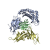
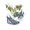
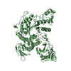
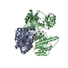
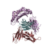
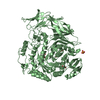
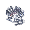
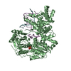
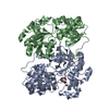
| Title | Crystal structure of HLA-A2 in complex with LILRB1 | ||||||
 Components Components |
| ||||||
 Keywords Keywords | IMMUNE SYSTEM / LILRB1 / peptide-MHC complex / HLA-A2 / peptide conformation / crystallisation chaperone | ||||||
| Function / homology |  Function and homology information Function and homology informationHLA-A specific inhibitory MHC class I receptor activity / : / negative regulation of alpha-beta T cell activation / negative regulation of serotonin secretion / MHC class Ib protein complex binding / HLA-B specific inhibitory MHC class I receptor activity / inhibitory MHC class I receptor activity / negative regulation of dendritic cell differentiation / Fc receptor mediated inhibitory signaling pathway / MHC class Ib receptor activity ...HLA-A specific inhibitory MHC class I receptor activity / : / negative regulation of alpha-beta T cell activation / negative regulation of serotonin secretion / MHC class Ib protein complex binding / HLA-B specific inhibitory MHC class I receptor activity / inhibitory MHC class I receptor activity / negative regulation of dendritic cell differentiation / Fc receptor mediated inhibitory signaling pathway / MHC class Ib receptor activity / immune response-inhibiting cell surface receptor signaling pathway / MHC class Ib protein binding / negative regulation of T cell mediated cytotoxicity / negative regulation of CD8-positive, alpha-beta T cell activation / negative regulation of transforming growth factor beta production / MHC class I receptor activity / negative regulation of cytokine production involved in immune response / dendritic cell differentiation / interleukin-10-mediated signaling pathway / negative regulation of osteoclast development / negative regulation of interleukin-12 production / protein phosphatase 1 binding / negative regulation of T cell activation via T cell receptor contact with antigen bound to MHC molecule on antigen presenting cell / negative regulation of dendritic cell apoptotic process / negative regulation of interferon-beta production / negative regulation of endocytosis / integrase activity / negative regulation of mononuclear cell proliferation / negative regulation of natural killer cell mediated cytotoxicity / positive regulation of macrophage cytokine production / Integration of viral DNA into host genomic DNA / Autointegration results in viral DNA circles / T cell proliferation involved in immune response / positive regulation of memory T cell activation / T cell mediated cytotoxicity directed against tumor cell target / positive regulation of CD8-positive, alpha-beta T cell activation / CD8-positive, alpha-beta T cell activation / positive regulation of CD8-positive, alpha-beta T cell proliferation / negative regulation of interleukin-10 production / Minus-strand DNA synthesis / Plus-strand DNA synthesis / Uncoating of the HIV Virion / 2-LTR circle formation / antigen processing and presentation of endogenous peptide antigen via MHC class I via ER pathway, TAP-dependent / TAP complex binding / Vpr-mediated nuclear import of PICs / antigen processing and presentation of exogenous peptide antigen via MHC class I / Golgi medial cisterna / Early Phase of HIV Life Cycle / Integration of provirus / APOBEC3G mediated resistance to HIV-1 infection / CD8 receptor binding / negative regulation of type II interferon production / negative regulation of cell cycle / negative regulation of calcium ion transport / protection from natural killer cell mediated cytotoxicity / endoplasmic reticulum exit site / TAP binding / beta-2-microglobulin binding / negative regulation of tumor necrosis factor production / MHC class I protein binding / Binding and entry of HIV virion / detection of bacterium / antigen processing and presentation of endogenous peptide antigen via MHC class Ib / antigen processing and presentation of endogenous peptide antigen via MHC class I via ER pathway, TAP-independent / negative regulation of T cell proliferation / T cell receptor binding / viral life cycle / positive regulation of defense response to virus by host / SH2 domain binding / early endosome lumen / Nef mediated downregulation of MHC class I complex cell surface expression / DAP12 interactions / T cell mediated cytotoxicity / Endosomal/Vacuolar pathway / HIV-1 retropepsin / symbiont-mediated activation of host apoptosis / retroviral ribonuclease H / exoribonuclease H / Antigen Presentation: Folding, assembly and peptide loading of class I MHC / lumenal side of endoplasmic reticulum membrane / regulation of iron ion transport / cellular response to iron(III) ion / antigen processing and presentation of exogenous protein antigen via MHC class Ib, TAP-dependent / exoribonuclease H activity / negative regulation of iron ion transport / negative regulation of forebrain neuron differentiation / regulation of erythrocyte differentiation / peptide antigen assembly with MHC class I protein complex / ER to Golgi transport vesicle membrane / response to molecule of bacterial origin / HFE-transferrin receptor complex / MHC class I peptide loading complex / transferrin transport / receptor internalization / negative regulation of receptor-mediated endocytosis / cellular response to iron ion / DNA integration / positive regulation of T cell cytokine production / Assembly Of The HIV Virion Similarity search - Function | ||||||
| Biological species |  Homo sapiens (human) Homo sapiens (human)  Human immunodeficiency virus 1 Human immunodeficiency virus 1 | ||||||
| Method |  X-RAY DIFFRACTION / SYNCHROTRON / MOLECULAR REPLACEMENT / Resolution: 2.39 Å X-RAY DIFFRACTION / SYNCHROTRON / MOLECULAR REPLACEMENT / Resolution: 2.39 Å | ||||||
| Model details | ================CATEGORY 5: Authors of Structure============================ Enter authors of the ...================CATEGORY 5: Authors of Structure============================ Enter authors of the deposited structures (e.g. Surname, F.M.) | ||||||
 Authors Authors | Stones, D.H. / Willcox, B.E. / Mohammed, F. | ||||||
 Citation Citation | Journal: J. Immunol. Methods / Year: 2019 Title: Application of the immunoregulatory receptor LILRB1 as a crystallisation chaperone for human class I MHC complexes. Authors: Mohammed, F. / Stones, D.H. / Willcox, B.E. | ||||||
| History |
|
- Structure visualization
| Structure viewer | Molecule: MolmilJmol/JSmol |
|---|
- Downloads & links
-Download
| PDBx/mmCIF format | 6ewa.cif.gz | 235.7 KB | Display | PDBx/mmCIF format |
|---|---|---|---|---|
| PDB format | pdb6ewa.ent.gz | 187.8 KB | Display | PDB format |
| PDBx/mmJSON format | 6ewa.json.gz | Tree view | PDBx/mmJSON format | |
| Others |  Other downloads Other downloads |
-Validation report
| Arichive directory | https://data.pdbj.org/pub/pdb/validation_reports/ew/6ewaftp://data.pdbj.org/pub/pdb/validation_reports/ew/6ewa | HTTPS FTP |
|---|
-Related structure data
| Related structure data |  6ewcC  6ewoC  1p7qS S: Starting model for refinement C: citing same article ( |
|---|---|
| Similar structure data |

-Links
 PDBj
PDBj


- Assembly
| Deposited unit | 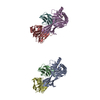
| ||||||||
|---|---|---|---|---|---|---|---|---|---|
| 1 | 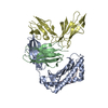
| ||||||||
| 2 | 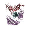
| ||||||||
| Unit cell |
|
-Components
| #1: Protein | Mass: 31951.316 Da / Num. of mol.: 2 Source method: isolated from a genetically manipulated source Source: (gene. exp.) Homo sapiens (human) / Gene: HLA-A, HLAA / Plasmid: pGMT7 / Production host:  #2: Protein | Mass: 11748.160 Da / Num. of mol.: 2 Source method: isolated from a genetically manipulated source Source: (gene. exp.) Homo sapiens (human) / Gene: B2M, CDABP0092, HDCMA22P / Plasmid: pGMT7 / Production host: #3: Protein/peptide | Mass: 993.199 Da / Num. of mol.: 2 / Source method: obtained synthetically / Source: (synth.) Human immunodeficiency virus 1 / References: UniProt: Q9WGX1, UniProt: P04585*PLUS#4: Protein | Mass: 21747.340 Da / Num. of mol.: 2 Source method: isolated from a genetically manipulated source Source: (gene. exp.) Homo sapiens (human) / Gene: LILRB1 / Plasmid: pET23a / Production host: #5: Water | ChemComp-HOH / |  Mass: 18.015 Da / Num. of mol.: 45 / Source method: isolated from a natural source / Formula: H2O Mass: 18.015 Da / Num. of mol.: 45 / Source method: isolated from a natural source / Formula: H2OHas protein modification | Y | |
|---|
-Experimental details
-Experiment
| Experiment | Method: X-RAY DIFFRACTION / Number of used crystals: 1 |
|---|
- Sample preparation
| Crystal | Density Matthews: 2.83 Å3/Da / Density % sol: 56.47 % |
|---|---|
| Crystal grow | Temperature: 298 K / Method: vapor diffusion, hanging drop Details: 17.5% PEG 3350, 0.2M Ammonium acetate, 0.1M Tris HCL |
-Data collection
| Diffraction | Mean temperature: 100 K | |||||||||||||||||||||||||||||||||||||||||||||||||||||||||||||||||||||||||||||||||||||||||||||||||||
|---|---|---|---|---|---|---|---|---|---|---|---|---|---|---|---|---|---|---|---|---|---|---|---|---|---|---|---|---|---|---|---|---|---|---|---|---|---|---|---|---|---|---|---|---|---|---|---|---|---|---|---|---|---|---|---|---|---|---|---|---|---|---|---|---|---|---|---|---|---|---|---|---|---|---|---|---|---|---|---|---|---|---|---|---|---|---|---|---|---|---|---|---|---|---|---|---|---|---|---|---|
| Diffraction source | Source: SYNCHROTRON / Site: ESRF  / Beamline: ID14-4 / Wavelength: 0.93927 Å / Beamline: ID14-4 / Wavelength: 0.93927 Å | |||||||||||||||||||||||||||||||||||||||||||||||||||||||||||||||||||||||||||||||||||||||||||||||||||
| Detector | Type: ADSC QUANTUM 4 / Detector: CCD / Date: Nov 8, 2006 | |||||||||||||||||||||||||||||||||||||||||||||||||||||||||||||||||||||||||||||||||||||||||||||||||||
| Radiation | Monochromator: mirrors / Protocol: SINGLE WAVELENGTH / Monochromatic (M) / Laue (L): M / Scattering type: x-ray | |||||||||||||||||||||||||||||||||||||||||||||||||||||||||||||||||||||||||||||||||||||||||||||||||||
| Radiation wavelength | Wavelength: 0.93927 Å / Relative weight: 1 | |||||||||||||||||||||||||||||||||||||||||||||||||||||||||||||||||||||||||||||||||||||||||||||||||||
| Reflection | Resolution: 2.39→48.679 Å / Num. obs: 60013 / % possible obs: 99.6 % / Redundancy: 9.6 % / Rmerge(I) obs: 0.122 / Rpim(I) all: 0.042 / Rrim(I) all: 0.13 / Rsym value: 0.122 / Net I/av σ(I): 5.2 / Net I/σ(I): 17.1 | |||||||||||||||||||||||||||||||||||||||||||||||||||||||||||||||||||||||||||||||||||||||||||||||||||
| Reflection shell | Diffraction-ID: 1
|
- Processing
| Software |
| ||||||||||||||||||||||||||||||||||||||||||||||||||||||||||||
|---|---|---|---|---|---|---|---|---|---|---|---|---|---|---|---|---|---|---|---|---|---|---|---|---|---|---|---|---|---|---|---|---|---|---|---|---|---|---|---|---|---|---|---|---|---|---|---|---|---|---|---|---|---|---|---|---|---|---|---|---|---|
| Refinement | Method to determine structure: MOLECULAR REPLACEMENT Starting model: 1P7Q Resolution: 2.39→44.59 Å / Cor.coef. Fo:Fc: 0.919 / Cor.coef. Fo:Fc free: 0.881 / SU B: 9.434 / SU ML: 0.214 / SU R Cruickshank DPI: 0.3572 / Cross valid method: THROUGHOUT / σ(F): 0 / ESU R: 0.357 / ESU R Free: 0.268 Details: HYDROGENS HAVE BEEN ADDED IN THE RIDING POSITIONS U VALUES : REFINED INDIVIDUALLY
| ||||||||||||||||||||||||||||||||||||||||||||||||||||||||||||
| Solvent computation | Ion probe radii: 0.8 Å / Shrinkage radii: 0.8 Å / VDW probe radii: 1.2 Å | ||||||||||||||||||||||||||||||||||||||||||||||||||||||||||||
| Displacement parameters | Biso max: 110.82 Å2 / Biso mean: 35.594 Å2 / Biso min: 14.51 Å2
| ||||||||||||||||||||||||||||||||||||||||||||||||||||||||||||
| Refinement step | Cycle: final / Resolution: 2.39→44.59 Å
| ||||||||||||||||||||||||||||||||||||||||||||||||||||||||||||
| Refine LS restraints |
| ||||||||||||||||||||||||||||||||||||||||||||||||||||||||||||
| LS refinement shell | Resolution: 2.392→2.454 Å / Rfactor Rfree error: 0 / Total num. of bins used: 20
|
