 Movie
Movie Controller

[English] 日本語
 Yorodumi
- PDB-6g4q: Structure of human ADP-forming succinyl-CoA ligase complex SUCLG1... -
+ Open data

- Basic information
| Entry | Database: PDB / ID: 6g4q | ||||||
|---|---|---|---|---|---|---|---|
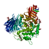
| Title | Structure of human ADP-forming succinyl-CoA ligase complex SUCLG1-SUCLA2 | ||||||
 Components Components |
| ||||||
 Keywords Keywords | LIGASE / Complex / Succinyl-CoA Ligase / Mitochondrial / ATP-specific | ||||||
| Function / homology |  Function and homology information Function and homology informationsuccinyl-CoA pathway / succinate-CoA ligase activity / malate-CoA ligase / succinate-CoA ligase complex (GDP-forming) / succinate-CoA ligase (GDP-forming) / succinate-CoA ligase (GDP-forming) activity / succinate-CoA ligase complex (ADP-forming) / succinate-CoA ligase (ADP-forming) / succinate-CoA ligase complex / succinate-CoA ligase (ADP-forming) activity ...succinyl-CoA pathway / succinate-CoA ligase activity / malate-CoA ligase / succinate-CoA ligase complex (GDP-forming) / succinate-CoA ligase (GDP-forming) / succinate-CoA ligase (GDP-forming) activity / succinate-CoA ligase complex (ADP-forming) / succinate-CoA ligase (ADP-forming) / succinate-CoA ligase complex / succinate-CoA ligase (ADP-forming) activity / Ligases; Forming carbon-sulfur bonds; Acid-thiol ligases / succinyl-CoA catabolic process / succinyl-CoA metabolic process / succinate metabolic process / Citric acid cycle (TCA cycle) / tricarboxylic acid cycle / mitochondrial matrix / nucleotide binding / magnesium ion binding / mitochondrion / RNA binding / extracellular exosome / ATP binding Similarity search - Function | ||||||
| Biological species |  Homo sapiens (human) Homo sapiens (human) | ||||||
| Method |  X-RAY DIFFRACTION / SYNCHROTRON / MOLECULAR REPLACEMENT / Resolution: 2.59 Å X-RAY DIFFRACTION / SYNCHROTRON / MOLECULAR REPLACEMENT / Resolution: 2.59 Å | ||||||
 Authors Authors | Bailey, H.J. / Shrestha, L. / Rembeza, E. / Sorrell, F.J. / Newman, J. / Strain-Damerell, C. / Burgess-Brown, N. / von Delft, F. / Arrowsmith, C. / Edwards, A. ...Bailey, H.J. / Shrestha, L. / Rembeza, E. / Sorrell, F.J. / Newman, J. / Strain-Damerell, C. / Burgess-Brown, N. / von Delft, F. / Arrowsmith, C. / Edwards, A. / Bountra, C. / Yue, W.W. | ||||||
 Citation Citation | Journal: To Be Published Title: Structure of human ADP-forming succinyl-CoA ligase complex SUCLG1-SUCLA2 Authors: Bailey, H.J. / Shrestha, L. / Rembeza, E. / Sorrell, F.J. / Strain-Damerell, C. / Burgess-Brown, N. / Arrowsmith, C. / Edwards, A. / Bountra, C. / Yue, W.W. | ||||||
| History |
|
- Structure visualization
| Structure viewer | Molecule: MolmilJmol/JSmol |
|---|
- Downloads & links
-Download
| PDBx/mmCIF format | 6g4q.cif.gz | 276.3 KB | Display | PDBx/mmCIF format |
|---|---|---|---|---|
| PDB format | pdb6g4q.ent.gz | 220.9 KB | Display | PDB format |
| PDBx/mmJSON format | 6g4q.json.gz | Tree view | PDBx/mmJSON format | |
| Others |  Other downloads Other downloads |
-Validation report
| Arichive directory | https://data.pdbj.org/pub/pdb/validation_reports/g4/6g4qftp://data.pdbj.org/pub/pdb/validation_reports/g4/6g4q | HTTPS FTP |
|---|
-Related structure data
| Related structure data |  1eucS S: Starting model for refinement |
|---|---|
| Similar structure data |


-Links
 PDBj
PDBj


- Assembly
| Deposited unit | 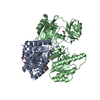
| ||||||||
|---|---|---|---|---|---|---|---|---|---|
| 1 |
| ||||||||
| Unit cell |
|
-Components
| #1: Protein | Mass: 32459.123 Da / Num. of mol.: 1 Source method: isolated from a genetically manipulated source Source: (gene. exp.) Homo sapiens (human) / Gene: SUCLG1 / Production host:  References: UniProt: P53597, succinate-CoA ligase (GDP-forming), succinate-CoA ligase (ADP-forming) | ||
|---|---|---|---|
| #2: Protein | Mass: 45198.273 Da / Num. of mol.: 1 Source method: isolated from a genetically manipulated source Source: (gene. exp.) Homo sapiens (human) / Gene: SUCLA2 / Production host: References: UniProt: Q9P2R7, succinate-CoA ligase (ADP-forming) | ||
| #3: Chemical | ChemComp-EDO /   Mass: 62.068 Da / Num. of mol.: 8 / Source method: obtained synthetically / Formula: C2H6O2 Mass: 62.068 Da / Num. of mol.: 8 / Source method: obtained synthetically / Formula: C2H6O2#4: Water | ChemComp-HOH / |  Mass: 18.015 Da / Num. of mol.: 82 / Source method: isolated from a natural source / Formula: H2O Mass: 18.015 Da / Num. of mol.: 82 / Source method: isolated from a natural source / Formula: H2O |
-Experimental details
-Experiment
| Experiment | Method: X-RAY DIFFRACTION / Number of used crystals: 1 |
|---|
- Sample preparation
| Crystal | Density Matthews: 3.78 Å3/Da / Density % sol: 67.44 % |
|---|---|
| Crystal grow | Temperature: 293.15 K / Method: vapor diffusion, sitting drop / pH: 7.5 / Details: 2.1M DL- malic acid |
-Data collection
| Diffraction | Mean temperature: 100 K |
|---|---|
| Diffraction source | Source: SYNCHROTRON / Site: Diamond  / Beamline: I04 / Wavelength: 0.9795 Å / Beamline: I04 / Wavelength: 0.9795 Å |
| Detector | Type: DECTRIS PILATUS 6M-F / Detector: PIXEL / Date: Nov 29, 2017 |
| Radiation | Protocol: SINGLE WAVELENGTH / Monochromatic (M) / Laue (L): M / Scattering type: x-ray |
| Radiation wavelength | Wavelength: 0.9795 Å / Relative weight: 1 |
| Reflection | Resolution: 2.59→65.1 Å / Num. obs: 36946 / % possible obs: 100 % / Redundancy: 9.7 % / CC1/2: 0.999 / Net I/σ(I): 14.4 |
| Reflection shell | Resolution: 2.59→2.66 Å / Mean I/σ(I) obs: 2.1 / Num. unique obs: 2712 / CC1/2: 0.547 / % possible all: 100 |
- Processing
| Software |
| ||||||||||||||||||||||||||||||||||||||||||||||||||||||||||||||||||||||||||||||||||||||||||||||||||||||||||||||||||||||||||||||||||||||||||||||||||||||||||||||||||||||||||||||||||||||||||||||||||||||||||||||||||||||||||||||||||||||||||||||||||||||||||
|---|---|---|---|---|---|---|---|---|---|---|---|---|---|---|---|---|---|---|---|---|---|---|---|---|---|---|---|---|---|---|---|---|---|---|---|---|---|---|---|---|---|---|---|---|---|---|---|---|---|---|---|---|---|---|---|---|---|---|---|---|---|---|---|---|---|---|---|---|---|---|---|---|---|---|---|---|---|---|---|---|---|---|---|---|---|---|---|---|---|---|---|---|---|---|---|---|---|---|---|---|---|---|---|---|---|---|---|---|---|---|---|---|---|---|---|---|---|---|---|---|---|---|---|---|---|---|---|---|---|---|---|---|---|---|---|---|---|---|---|---|---|---|---|---|---|---|---|---|---|---|---|---|---|---|---|---|---|---|---|---|---|---|---|---|---|---|---|---|---|---|---|---|---|---|---|---|---|---|---|---|---|---|---|---|---|---|---|---|---|---|---|---|---|---|---|---|---|---|---|---|---|---|---|---|---|---|---|---|---|---|---|---|---|---|---|---|---|---|---|---|---|---|---|---|---|---|---|---|---|---|---|---|---|---|---|---|---|---|---|---|---|---|---|---|---|---|---|---|---|---|---|
| Refinement | Method to determine structure: MOLECULAR REPLACEMENT Starting model: 1EUC Resolution: 2.59→65.1 Å / SU ML: 0.42 / Cross valid method: THROUGHOUT / σ(F): 1.33 / Phase error: 31.31
| ||||||||||||||||||||||||||||||||||||||||||||||||||||||||||||||||||||||||||||||||||||||||||||||||||||||||||||||||||||||||||||||||||||||||||||||||||||||||||||||||||||||||||||||||||||||||||||||||||||||||||||||||||||||||||||||||||||||||||||||||||||||||||
| Solvent computation | Shrinkage radii: 0.9 Å / VDW probe radii: 1.11 Å | ||||||||||||||||||||||||||||||||||||||||||||||||||||||||||||||||||||||||||||||||||||||||||||||||||||||||||||||||||||||||||||||||||||||||||||||||||||||||||||||||||||||||||||||||||||||||||||||||||||||||||||||||||||||||||||||||||||||||||||||||||||||||||
| Displacement parameters | Biso max: 187.17 Å2 / Biso mean: 78.9871 Å2 / Biso min: 37.29 Å2 | ||||||||||||||||||||||||||||||||||||||||||||||||||||||||||||||||||||||||||||||||||||||||||||||||||||||||||||||||||||||||||||||||||||||||||||||||||||||||||||||||||||||||||||||||||||||||||||||||||||||||||||||||||||||||||||||||||||||||||||||||||||||||||
| Refinement step | Cycle: final / Resolution: 2.59→65.1 Å
| ||||||||||||||||||||||||||||||||||||||||||||||||||||||||||||||||||||||||||||||||||||||||||||||||||||||||||||||||||||||||||||||||||||||||||||||||||||||||||||||||||||||||||||||||||||||||||||||||||||||||||||||||||||||||||||||||||||||||||||||||||||||||||
| LS refinement shell | Refine-ID: X-RAY DIFFRACTION / Rfactor Rfree error: 0 / Total num. of bins used: 12
| ||||||||||||||||||||||||||||||||||||||||||||||||||||||||||||||||||||||||||||||||||||||||||||||||||||||||||||||||||||||||||||||||||||||||||||||||||||||||||||||||||||||||||||||||||||||||||||||||||||||||||||||||||||||||||||||||||||||||||||||||||||||||||
| Refinement TLS params. | Method: refined / Refine-ID: X-RAY DIFFRACTION
| ||||||||||||||||||||||||||||||||||||||||||||||||||||||||||||||||||||||||||||||||||||||||||||||||||||||||||||||||||||||||||||||||||||||||||||||||||||||||||||||||||||||||||||||||||||||||||||||||||||||||||||||||||||||||||||||||||||||||||||||||||||||||||
| Refinement TLS group |
|
