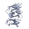
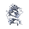
登録情報 データベース : PDB / ID : 6ew1タイトル Crystal structure of the Filamin A Ig-like domains 3-5 mutant P637Q Filamin-A キーワード / 機能・相同性 分子機能 ドメイン・相同性 構成要素
/ / / / / / / / / / / / / / / / / / / / / / / / / / / / / / / / / / / / / / / / / / / / / / / / / / / / / / / / / / / / / / / / / / / / / / / / / / / / / / / / / / / / / / / / / / / / / / / / / / / / / / / / / / / / / / / / / / / / / 生物種 Homo sapiens (ヒト)手法 / / / 解像度 : 2.3070081803 Å データ登録者 Haataja, T.J.K. / Pentikainen, U. 資金援助 組織 認可番号 国 Academy of Finland 283481
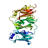
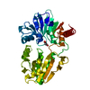
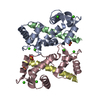
ジャーナル : Structure / 年 : 2019タイトル : Non-syndromic Mitral Valve Dysplasia Mutation Changes the Force Resilience and Interaction of Human Filamin A.著者 : Tatu J K Haataja / Rafael C Bernardi / Simon Lecointe / Romain Capoulade / Jean Merot / Ulla Pentikäinen / 要旨 : Filamin A (FLNa), expressed in endocardial endothelia during fetal valve morphogenesis, is key in cardiac development. Missense mutations in FLNa cause non-syndromic mitral valve dysplasia (FLNA-MVD). ... Filamin A (FLNa), expressed in endocardial endothelia during fetal valve morphogenesis, is key in cardiac development. Missense mutations in FLNa cause non-syndromic mitral valve dysplasia (FLNA-MVD). Here, we aimed to reveal the currently unknown underlying molecular mechanism behind FLNA-MVD caused by the FLNa P637Q mutation. The solved crystal structure of the FLNa3-5 P637Q revealed that this mutation causes only minor structural changes close to mutation site. These changes were observed to significantly affect FLNa's ability to transmit cellular force and to interact with its binding partner. The performed steered molecular dynamics simulations showed that significantly lower forces are needed to split domains 4 and 5 in FLNA-MVD than with wild-type FLNa. The P637Q mutation was also observed to interfere with FLNa's interactions with the protein tyrosine phosphatase PTPN12. Our results provide a crucial step toward understanding the molecular bases behind FLNA-MVD, which is critical for the development of drug-based therapeutics. 履歴 登録 2017年11月3日 登録サイト / 処理サイト 改定 1.0 2018年10月31日 Provider / タイプ 改定 1.1 2019年1月16日 Group / Database references / カテゴリ Item _citation.journal_id_ISSN / _citation.journal_volume ... _citation.journal_id_ISSN / _citation.journal_volume / _citation.page_first / _citation.page_last / _citation.year 改定 1.2 2024年1月17日 Group Advisory / Data collection ... Advisory / Data collection / Database references / Refinement description カテゴリ chem_comp_atom / chem_comp_bond ... chem_comp_atom / chem_comp_bond / database_2 / pdbx_initial_refinement_model / pdbx_unobs_or_zero_occ_atoms Item / _database_2.pdbx_database_accession
すべて表示 表示を減らす
 ムービー
ムービー コントローラー


 データを開く
 基本情報
基本情報 要素
要素 キーワード
キーワード 機能・相同性情報
 Homo sapiens (ヒト)
 X線回折 /
  データ登録者
 フィンランド, 1件
  引用
引用

 構造の表示
構造の表示 ダウンロードとリンク
 その他のダウンロード


 PDBj
PDBj


 集合体
集合体

 分子量: 18.015 Da / 分子数: 53 / 由来タイプ: 天然 / 式: H2O
 試料調製
試料調製 解析
解析