[English] 日本語
Yorodumi
- SASDEP7: Filamin A Ig-like domains 3-5 P637Q mutant (FLNa3-5 P637Q) -

+
Open data


ID or keywords:

Loading...

-
Basic information

Entry
Database: SASBDB / ID: SASDEP7
SampleFilamin A Ig-like domains 3-5 P637Q mutant (FLNa3-5 P637Q)
  • Filamin A Ig.like domains 3-5 P637Q mutant (protein), FLNa3-5 P637Q, Homo sapiens
Function / homology
Function and homology information


regulation of membrane repolarization during atrial cardiac muscle cell action potential / regulation of membrane repolarization during cardiac muscle cell action potential / establishment of Sertoli cell barrier / Myb complex / glycoprotein Ib-IX-V complex / adenylate cyclase-inhibiting dopamine receptor signaling pathway / formation of radial glial scaffolds / positive regulation of integrin-mediated signaling pathway / actin crosslink formation / tubulin deacetylation ...regulation of membrane repolarization during atrial cardiac muscle cell action potential / regulation of membrane repolarization during cardiac muscle cell action potential / establishment of Sertoli cell barrier / Myb complex / glycoprotein Ib-IX-V complex / adenylate cyclase-inhibiting dopamine receptor signaling pathway / formation of radial glial scaffolds / positive regulation of integrin-mediated signaling pathway / actin crosslink formation / tubulin deacetylation / blood coagulation, intrinsic pathway / protein localization to bicellular tight junction / OAS antiviral response / positive regulation of actin filament bundle assembly / positive regulation of neuron migration / Cell-extracellular matrix interactions / early endosome to late endosome transport / Fc-gamma receptor I complex binding / positive regulation of potassium ion transmembrane transport / apical dendrite / cell-cell junction organization / positive regulation of neural precursor cell proliferation / positive regulation of platelet activation / protein localization to cell surface / wound healing, spreading of cells / podosome / negative regulation of transcription by RNA polymerase I / megakaryocyte development / GP1b-IX-V activation signalling / receptor clustering / positive regulation of axon regeneration / cortical cytoskeleton / SMAD binding / RHO GTPases activate PAKs / actin filament bundle / brush border / semaphorin-plexin signaling pathway / cilium assembly / blood vessel remodeling / mitotic spindle assembly / epithelial to mesenchymal transition / potassium channel regulator activity / axonal growth cone / heart morphogenesis / release of sequestered calcium ion into cytosol / positive regulation of substrate adhesion-dependent cell spreading / protein sequestering activity / regulation of cell migration / dendritic shaft / protein kinase C binding / protein localization to plasma membrane / actin filament / negative regulation of DNA-binding transcription factor activity / trans-Golgi network / mRNA transcription by RNA polymerase II / G protein-coupled receptor binding / establishment of protein localization / synapse organization / negative regulation of protein catabolic process / cerebral cortex development / small GTPase binding / Z disc / kinase binding / platelet aggregation / positive regulation of protein import into nucleus / actin filament binding / cell-cell junction / Platelet degranulation / actin cytoskeleton / negative regulation of neuron projection development / actin cytoskeleton organization / GTPase binding / angiogenesis / DNA-binding transcription factor binding / perikaryon / transmembrane transporter binding / positive regulation of canonical NF-kappaB signal transduction / postsynapse / protein stabilization / cadherin binding / focal adhesion / negative regulation of apoptotic process / nucleolus / perinuclear region of cytoplasm / glutamatergic synapse / protein homodimerization activity / RNA binding / extracellular exosome / extracellular region / nucleus / membrane / plasma membrane / cytosol / cytoplasm
Similarity search - Function
Filamin family / Filamin/ABP280 repeat / Filamin-type immunoglobulin domains / Filamin/ABP280 repeat / Filamin/ABP280 repeat profile. / Filamin/ABP280 repeat-like / Actinin-type actin-binding domain signature 1. / Actinin-type actin-binding domain signature 2. / Actinin-type actin-binding domain, conserved site / Calponin homology domain ...Filamin family / Filamin/ABP280 repeat / Filamin-type immunoglobulin domains / Filamin/ABP280 repeat / Filamin/ABP280 repeat profile. / Filamin/ABP280 repeat-like / Actinin-type actin-binding domain signature 1. / Actinin-type actin-binding domain signature 2. / Actinin-type actin-binding domain, conserved site / Calponin homology domain / Calponin homology (CH) domain / Calponin homology domain / CH domain superfamily / Calponin homology (CH) domain profile. / Immunoglobulin E-set / Immunoglobulin-like fold
Similarity search - Domain/homology
Biological speciesHomo sapiens (human)
Citation
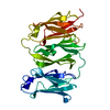
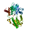
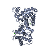
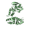
Journal: Structure / Year: 2019
Title: Non-syndromic Mitral Valve Dysplasia Mutation Changes the Force Resilience and Interaction of Human Filamin A.
Authors: Tatu J K Haataja / Rafael C Bernardi / Simon Lecointe / Romain Capoulade / Jean Merot / Ulla Pentikäinen /
Abstract: Filamin A (FLNa), expressed in endocardial endothelia during fetal valve morphogenesis, is key in cardiac development. Missense mutations in FLNa cause non-syndromic mitral valve dysplasia (FLNA-MVD). ...Filamin A (FLNa), expressed in endocardial endothelia during fetal valve morphogenesis, is key in cardiac development. Missense mutations in FLNa cause non-syndromic mitral valve dysplasia (FLNA-MVD). Here, we aimed to reveal the currently unknown underlying molecular mechanism behind FLNA-MVD caused by the FLNa P637Q mutation. The solved crystal structure of the FLNa3-5 P637Q revealed that this mutation causes only minor structural changes close to mutation site. These changes were observed to significantly affect FLNa's ability to transmit cellular force and to interact with its binding partner. The performed steered molecular dynamics simulations showed that significantly lower forces are needed to split domains 4 and 5 in FLNA-MVD than with wild-type FLNa. The P637Q mutation was also observed to interfere with FLNa's interactions with the protein tyrosine phosphatase PTPN12. Our results provide a crucial step toward understanding the molecular bases behind FLNA-MVD, which is critical for the development of drug-based therapeutics.
Contact author
  • Tatu Haataja (University of Jyväskylä, Jyväskylän, Finland)

-
Structure visualization

Structure viewerMolecule:
MolmilJmol/JSmol

Downloads & links

-
Models

Model #2607
Type: atomic / Chi-square value: 1.123
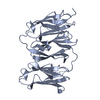
Search similar-shape structures of this assembly by Omokage search (details)

-
Sample

SampleName: Filamin A Ig-like domains 3-5 P637Q mutant (FLNa3-5 P637Q)
Specimen concentration: 1.00-4.00
BufferName: 20 mM Tris, 100 mM NaCl, 1 mM DTT / pH: 8
Entity #850Name: FLNa3-5 P637Q / Type: protein / Description: Filamin A Ig.like domains 3-5 P637Q mutant / Formula weight: 30.791 / Num. of mol.: 1 / Source: Homo sapiens / References: UniProt: P21333
Sequence: CNPSACRAVG RGLQPKGVRV KETADFKVYT KGAGSGELKV TVKGPKGEER VKQKDLGDGV YGFEYYPMVP GTYIVTITWG GQNIGRSPFE VKVGTECGNQ KVRAWGPGLE GGVVGKSADF VVEAIGDDVG TLGFSVEGPS QAKIECDDKG DGSCDVRYWQ QEAGEYAVHV ...Sequence:
CNPSACRAVG RGLQPKGVRV KETADFKVYT KGAGSGELKV TVKGPKGEER VKQKDLGDGV YGFEYYPMVP GTYIVTITWG GQNIGRSPFE VKVGTECGNQ KVRAWGPGLE GGVVGKSADF VVEAIGDDVG TLGFSVEGPS QAKIECDDKG DGSCDVRYWQ QEAGEYAVHV LCNSEDIRLS PFMADIRDAP QDFHPDRVKA RGPGLEKTGV AVNKPAEFTV DAKHGGKAPL RVQVQDNEGC PVEALVKDNG NGTYSCSYVP RKPVKHTAMV SWGGVSIPNS PFRVNVGAG

-
Experimental information

BeamInstrument name: ESRF BM29 / City: Grenoble / : France / Type of source: X-ray synchrotron / Wavelength: 0.1 Å / Dist. spec. to detc.: 2.9 mm
DetectorName: Pilatus 1M / Type: Dectris / Pixsize x: 172 mm
Scan
Title: Filamin A Ig-like domains 3-5 P637Q mutant (FLNa3-5 P637Q)
Measurement date: Feb 10, 2017 / Cell temperature: 20 °C / Exposure time: 1 sec. / Number of frames: 10 / Unit: 1/nm /
MinMax
Q0.0553 4.8334
Distance distribution function P(R)
Sofotware P(R): GNOM 4.6 / Number of points: 364 /
MinMax
Q0.08119 3.499
P(R) point6 369
R0 7.4
Result
Type of curve: merged
Comments: SAXS data describing the Filamin A Ig-like domains 3-5 P637Q mutant in solution. The experiment was done to enable comparison with the corresponding WT fragment and to validate the crystal ...Comments: SAXS data describing the Filamin A Ig-like domains 3-5 P637Q mutant in solution. The experiment was done to enable comparison with the corresponding WT fragment and to validate the crystal structure of the mutant (SASDEQ7).
ExperimentalPorod
MW23.24 kDa-
Volume-39 nm3

P(R)GuinierGuinier error
Forward scattering, I024.25 24.26 0.1
Radius of gyration, Rg2.21 nm2.21 nm0.1

MinMaxError
D-7.4 0.5
Guinier point3 113 -

+
About Yorodumi

-
News

-
Feb 9, 2022. New format data for meta-information of EMDB entries

New format data for meta-information of EMDB entries

  • Version 3 of the EMDB header file is now the official format.
  • The previous official version 1.9 will be removed from the archive.

Related info.:EMDB header

External links:wwPDB to switch to version 3 of the EMDB data model

-
Aug 12, 2020. Covid-19 info

Covid-19 info

URL: https://pdbj.org/emnavi/covid19.php

New page: Covid-19 featured information page in EM Navigator.

Related info.:Covid-19 info / Mar 5, 2020. Novel coronavirus structure data

+
Mar 5, 2020. Novel coronavirus structure data

Novel coronavirus structure data

Related info.:Yorodumi Speices / Aug 12, 2020. Covid-19 info

External links:COVID-19 featured content - PDBj / Molecule of the Month (242):Coronavirus Proteases

+
Jan 31, 2019. EMDB accession codes are about to change! (news from PDBe EMDB page)

EMDB accession codes are about to change! (news from PDBe EMDB page)

  • The allocation of 4 digits for EMDB accession codes will soon come to an end. Whilst these codes will remain in use, new EMDB accession codes will include an additional digit and will expand incrementally as the available range of codes is exhausted. The current 4-digit format prefixed with “EMD-” (i.e. EMD-XXXX) will advance to a 5-digit format (i.e. EMD-XXXXX), and so on. It is currently estimated that the 4-digit codes will be depleted around Spring 2019, at which point the 5-digit format will come into force.
  • The EM Navigator/Yorodumi systems omit the EMD- prefix.

Related info.:Q: What is EMD? / ID/Accession-code notation in Yorodumi/EM Navigator

External links:EMDB Accession Codes are Changing Soon! / Contact to PDBj

+
Jul 12, 2017. Major update of PDB

Major update of PDB

  • wwPDB released updated PDB data conforming to the new PDBx/mmCIF dictionary.
  • This is a major update changing the version number from 4 to 5, and with Remediation, in which all the entries are updated.
  • In this update, many items about electron microscopy experimental information are reorganized (e.g. em_software).
  • Now, EM Navigator and Yorodumi are based on the updated data.

External links:wwPDB Remediation / Enriched Model Files Conforming to OneDep Data Standards Now Available in the PDB FTP Archive

-
Yorodumi

Thousand views of thousand structures

  • Yorodumi is a browser for structure data from EMDB, PDB, SASBDB, etc.
  • This page is also the successor to EM Navigator detail page, and also detail information page/front-end page for Omokage search.
  • The word "yorodu" (or yorozu) is an old Japanese word meaning "ten thousand". "mi" (miru) is to see.

Related info.:EMDB / PDB / SASBDB / Comparison of 3 databanks / Yorodumi Search / Aug 31, 2016. New EM Navigator & Yorodumi / Yorodumi Papers / Jmol/JSmol / Function and homology information / Changes in new EM Navigator and Yorodumi

Read more