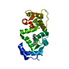
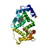
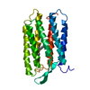
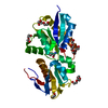
FI18190p1 / Frequenin 2 / isoform A / isoform B / isoform C / isoform D
Mass: 21922.443 Da / Num. of mol.: 1 Mutation: As reported in a previous publication (Romero-Pozuelo et al., JCS 2014) Drosophila Frq2 suffers edition process and besides de curated sequence, the I178M mutant also exists naturally Source method: isolated from a genetically manipulated source Source: (gene. exp.) Drosophila melanogaster (fruit fly) Gene: Frq2, Dmel\CG5907, Frq, frq, frq2, Frq2-RA, CG5907, Dmel_CG5907 Production host: Escherichia coli (E. coli) / References: UniProt: Q9VWX8
In the structure databanks used in Yorodumi, some data are registered as the other names, "COVID-19 virus" and "2019-nCoV". Here are the details of the virus and the list of structure data.
Jan 31, 2019. EMDB accession codes are about to change! (news from PDBe EMDB page)
EMDB accession codes are about to change! (news from PDBe EMDB page)
The allocation of 4 digits for EMDB accession codes will soon come to an end. Whilst these codes will remain in use, new EMDB accession codes will include an additional digit and will expand incrementally as the available range of codes is exhausted. The current 4-digit format prefixed with “EMD-” (i.e. EMD-XXXX) will advance to a 5-digit format (i.e. EMD-XXXXX), and so on. It is currently estimated that the 4-digit codes will be depleted around Spring 2019, at which point the 5-digit format will come into force.
The EM Navigator/Yorodumi systems omit the EMD- prefix.
Related info.:Q: What is EMD? / ID/Accession-code notation in Yorodumi/EM Navigator
Yorodumi is a browser for structure data from EMDB, PDB, SASBDB, etc.
This page is also the successor to EM Navigator detail page, and also detail information page/front-end page for Omokage search.
The word "yorodu" (or yorozu) is an old Japanese word meaning "ten thousand". "mi" (miru) is to see.
Related info.:EMDB / PDB / SASBDB / Comparison of 3 databanks / Yorodumi Search / Aug 31, 2016. New EM Navigator & Yorodumi / Yorodumi Papers / Jmol/JSmol / Function and homology information / Changes in new EM Navigator and Yorodumi
 Movie
Movie Controller

 Yorodumi
 Open data
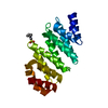
 Basic information
 Components
 Keywords
 Function and homology information
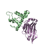
 X-RAY DIFFRACTION /
  Authors
 Spain, 4items
  Citation
 Structure visualization
 Downloads & links
 Other downloads
 PDBj
PDBj Assembly


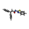


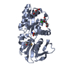
 Mass: 40.078 Da / Num. of mol.: 3 / Source method: obtained synthetically / Formula: Ca
 Mass: 358.456 Da / Num. of mol.: 1 / Source method: obtained synthetically / Formula: C22H18N2OS
 Mass: 120.147 Da / Num. of mol.: 1 / Source method: obtained synthetically / Formula: C5H12O3 / Comment: precipitant*YM
 Mass: 22.990 Da / Num. of mol.: 1 / Source method: obtained synthetically / Formula: Na
 Sample preparation
 Processing