 Movie
Movie Controller


+ Open data

- Basic information
| Entry | Database: PDB / ID: 1rfy | ||||||
|---|---|---|---|---|---|---|---|
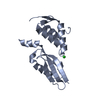
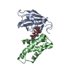
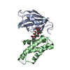
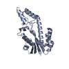
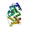
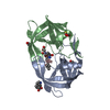
| Title | Crystal Structure of Quorum-Sensing Antiactivator TraM | ||||||
 Components Components | Transcriptional repressor traM | ||||||
 Keywords Keywords | TRANSCRIPTION / inter- and intra-molcular two-helix coiled coil / homodimer | ||||||
| Function / homology | Transcriptional repressor TraM / Transcriptional repressor TraM superfamily / Prokaryotic Transcriptional repressor TraM / HR1 repeat / Helix Hairpins / negative regulation of DNA-templated transcription / Orthogonal Bundle / Mainly Alpha / Transcriptional repressor TraM Function and homology information Function and homology information | ||||||
| Biological species |  Agrobacterium tumefaciens (bacteria) Agrobacterium tumefaciens (bacteria) | ||||||
| Method |  X-RAY DIFFRACTION / SYNCHROTRON / SAD / Resolution: 1.6 Å X-RAY DIFFRACTION / SYNCHROTRON / SAD / Resolution: 1.6 Å | ||||||
 Authors Authors | Chen, G. / Malenkos, J.W. / Cha, M.R. / Fuqua, C. / Chen, L. | ||||||
 Citation Citation | Journal: Mol.Microbiol. / Year: 2004 Title: Quorum-sensing antiactivator TraM forms a dimer that dissociates to inhibit TraR Authors: Chen, G. / Malenkos, J.W. / Cha, M.R. / Fuqua, C. / Chen, L. | ||||||
| History |
|
- Structure visualization
| Structure viewer | Molecule: MolmilJmol/JSmol |
|---|
- Downloads & links
-Download
| PDBx/mmCIF format | 1rfy.cif.gz | 49.6 KB | Display | PDBx/mmCIF format |
|---|---|---|---|---|
| PDB format | pdb1rfy.ent.gz | 36.3 KB | Display | PDB format |
| PDBx/mmJSON format | 1rfy.json.gz | Tree view | PDBx/mmJSON format | |
| Others |  Other downloads Other downloads |
-Validation report
| Arichive directory | https://data.pdbj.org/pub/pdb/validation_reports/rf/1rfyftp://data.pdbj.org/pub/pdb/validation_reports/rf/1rfy | HTTPS FTP |
|---|
-Related structure data
| Similar structure data |
|---|


-Links
 PDBj
PDBj- Assembly
| Deposited unit | 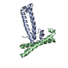
| ||||||||||
|---|---|---|---|---|---|---|---|---|---|---|---|
| 1 | 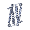
| ||||||||||
| 2 | 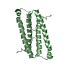
| ||||||||||
| Unit cell |
| ||||||||||
| Details | the second protomer of the biological assembly is generated by the two-fold axis: 2-x, 1-y, z the second protomer of the biological assembly is generated by the two-fold axis: 1.5-x, 0.5-y, z |
-Components
| #1: Protein | Mass: 11365.187 Da / Num. of mol.: 2 Source method: isolated from a genetically manipulated source Source: (gene. exp.) Agrobacterium tumefaciens (bacteria) / Gene: traM / Plasmid: pET15b / Production host: #2: Water | ChemComp-HOH / |  Mass: 18.015 Da / Num. of mol.: 230 / Source method: isolated from a natural source / Formula: H2O Mass: 18.015 Da / Num. of mol.: 230 / Source method: isolated from a natural source / Formula: H2O |
|---|
-Experimental details
-Experiment
| Experiment | Method: X-RAY DIFFRACTION / Number of used crystals: 1 |
|---|
- Sample preparation
| Crystal | Density Matthews: 2.54 Å3/Da / Density % sol: 51.2 % |
|---|---|
| Crystal grow | Temperature: 298 K / Method: vapor diffusion, hanging drop / pH: 5.6 Details: amonium acetate, sodium citrate, PEG 2000, pH 5.6, VAPOR DIFFUSION, HANGING DROP, temperature 298K |
-Data collection
| Diffraction | Mean temperature: 100 K |
|---|---|
| Diffraction source | Source: SYNCHROTRON / Site: CHESS  / Beamline: F2 / Wavelength: 0.9636 Å / Beamline: F2 / Wavelength: 0.9636 Å |
| Detector | Type: ADSC QUANTUM 4 / Detector: CCD / Date: Jan 17, 2003 |
| Radiation | Monochromator: Si (111) / Protocol: SINGLE WAVELENGTH / Monochromatic (M) / Laue (L): M / Scattering type: x-ray |
| Radiation wavelength | Wavelength: 0.9636 Å / Relative weight: 1 |
| Reflection | Resolution: 1.6→60 Å / Num. all: 30102 / Num. obs: 29690 / % possible obs: 98.5 % / Observed criterion σ(I): 1 / Redundancy: 9.9 % / Biso Wilson estimate: 21.3 Å2 / Rmerge(I) obs: 0.061 / Net I/σ(I): 37.4 |
| Reflection shell | Resolution: 1.6→1.63 Å / Rmerge(I) obs: 0.403 / Mean I/σ(I) obs: 4.1 / Num. unique all: 1234 / % possible all: 84.8 |
- Processing
| Software |
| ||||||||||||||||||||||||||||||||||||
|---|---|---|---|---|---|---|---|---|---|---|---|---|---|---|---|---|---|---|---|---|---|---|---|---|---|---|---|---|---|---|---|---|---|---|---|---|---|
| Refinement | Method to determine structure: SAD / Resolution: 1.6→60 Å / Rfactor Rfree error: 0.004 / Data cutoff high absF: 293645.01 / Data cutoff low absF: 0 / Isotropic thermal model: RESTRAINED / Cross valid method: THROUGHOUT / σ(F): 0 / Stereochemistry target values: Engh & Huber
| ||||||||||||||||||||||||||||||||||||
| Solvent computation | Solvent model: FLAT MODEL / Bsol: 52.2428 Å2 / ksol: 0.392037 e/Å3 | ||||||||||||||||||||||||||||||||||||
| Displacement parameters | Biso mean: 20.6 Å2
| ||||||||||||||||||||||||||||||||||||
| Refine analyze |
| ||||||||||||||||||||||||||||||||||||
| Refinement step | Cycle: LAST / Resolution: 1.6→60 Å
| ||||||||||||||||||||||||||||||||||||
| Refine LS restraints |
| ||||||||||||||||||||||||||||||||||||
| LS refinement shell | Resolution: 1.6→1.7 Å / Rfactor Rfree error: 0.011 / Total num. of bins used: 6
| ||||||||||||||||||||||||||||||||||||
| Xplor file |
|
