 Movie
Movie Controller


+ Open data

- Basic information
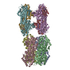
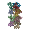
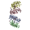
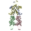
| Entry | Database: PDB / ID: 6e9x | ||||||
|---|---|---|---|---|---|---|---|
| Title | DHF91 filament | ||||||
 Components Components | DHF91 filament | ||||||
 Keywords Keywords | PROTEIN FIBRIL / Protein design / filament | ||||||
| Biological species | synthetic construct (others) | ||||||
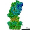
| Method | ELECTRON MICROSCOPY / helical reconstruction / cryo EM / Resolution: 7.8 Å | ||||||
 Authors Authors | Lynch, E.M. / Shen, H. / Fallas, J.A. / Kollman, J.M. / Baker, D. | ||||||
 Citation Citation | Journal: Science / Year: 2018 Title: De novo design of self-assembling helical protein filaments. Authors: Hao Shen / Jorge A Fallas / Eric Lynch / William Sheffler / Bradley Parry / Nicholas Jannetty / Justin Decarreau / Michael Wagenbach / Juan Jesus Vicente / Jiajun Chen / Lei Wang / Quinton ...Authors: Hao Shen / Jorge A Fallas / Eric Lynch / William Sheffler / Bradley Parry / Nicholas Jannetty / Justin Decarreau / Michael Wagenbach / Juan Jesus Vicente / Jiajun Chen / Lei Wang / Quinton Dowling / Gustav Oberdorfer / Lance Stewart / Linda Wordeman / James De Yoreo / Christine Jacobs-Wagner / Justin Kollman / David Baker /   Abstract: We describe a general computational approach to designing self-assembling helical filaments from monomeric proteins and use this approach to design proteins that assemble into micrometer-scale ...We describe a general computational approach to designing self-assembling helical filaments from monomeric proteins and use this approach to design proteins that assemble into micrometer-scale filaments with a wide range of geometries in vivo and in vitro. Cryo-electron microscopy structures of six designs are close to the computational design models. The filament building blocks are idealized repeat proteins, and thus the diameter of the filaments can be systematically tuned by varying the number of repeat units. The assembly and disassembly of the filaments can be controlled by engineered anchor and capping units built from monomers lacking one of the interaction surfaces. The ability to generate dynamic, highly ordered structures that span micrometers from protein monomers opens up possibilities for the fabrication of new multiscale metamaterials. | ||||||
| History |
|
- Structure visualization
| Movie |
 Movie viewer Movie viewer |
|---|---|
| Structure viewer | Molecule: MolmilJmol/JSmol |
- Downloads & links
-Download
| PDBx/mmCIF format | 6e9x.cif.gz | 1 MB | Display | PDBx/mmCIF format |
|---|---|---|---|---|
| PDB format | pdb6e9x.ent.gz | 891.3 KB | Display | PDB format |
| PDBx/mmJSON format | 6e9x.json.gz | Tree view | PDBx/mmJSON format | |
| Others |  Other downloads Other downloads |
-Validation report
| Summary document | 6e9x_validation.pdf.gz | 929.9 KB | Display | wwPDB validaton report |
|---|---|---|---|---|
| Full document | 6e9x_full_validation.pdf.gz | 1004.5 KB | Display | |
| Data in XML | 6e9x_validation.xml.gz | 90.5 KB | Display | |
| Data in CIF | 6e9x_validation.cif.gz | 127.3 KB | Display | |
| Arichive directory | https://data.pdbj.org/pub/pdb/validation_reports/e9/6e9xftp://data.pdbj.org/pub/pdb/validation_reports/e9/6e9x | HTTPS FTP |
-Related structure data
| Related structure data | 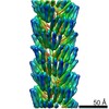 9019MC 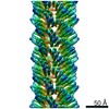 9016C  9017C 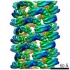 9018C 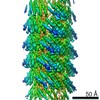 9020C 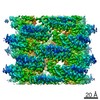 9021C 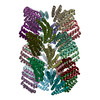 6e9rC 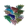 6e9tC 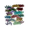 6e9vC 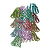 6e9yC 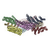 6e9zC M: map data used to model this data C: citing same article ( |
|---|---|
| Similar structure data |

-Links
 PDBj
PDBj
- Assembly
| Deposited unit | 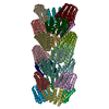
|
|---|---|
| 1 |
|
| Symmetry | Helical symmetry: (Circular symmetry: 3 / Dyad axis: no / N subunits divisor: 1 / Num. of operations: 7 / Rise per n subunits: 24.1338 Å / Rotation per n subunits: 50.1109 °) |
-Components
| #1: Protein | Mass: 17830.607 Da / Num. of mol.: 21 Source method: isolated from a genetically manipulated source Source: (gene. exp.) synthetic construct (others) / Production host:  |
|---|
-Experimental details
-Experiment
| Experiment | Method: ELECTRON MICROSCOPY |
|---|---|
| EM experiment | Aggregation state: FILAMENT / 3D reconstruction method: helical reconstruction |
- Sample preparation
| Component | Name: DHF91 filament / Type: COMPLEX / Entity ID: all / Source: RECOMBINANT |
|---|---|
| Source (natural) | Organism: synthetic construct (others) |
| Source (recombinant) | Organism: |
| Buffer solution | pH: 8 |
| Specimen | Embedding applied: NO / Shadowing applied: NO / Staining applied: NO / Vitrification applied: YES |
| Specimen support | Details: unspecified |
| Vitrification | Cryogen name: ETHANE |
- Electron microscopy imaging
| Experimental equipment |  Model: Tecnai F20 / Image courtesy: FEI Company |
|---|---|
| Microscopy | Model: FEI TECNAI F20 |
| Electron gun | Electron source:  FIELD EMISSION GUN / Accelerating voltage: 200 kV / Illumination mode: FLOOD BEAM FIELD EMISSION GUN / Accelerating voltage: 200 kV / Illumination mode: FLOOD BEAM |
| Electron lens | Mode: BRIGHT FIELD |
| Image recording | Electron dose: 45 e/Å2 / Detector mode: COUNTING / Film or detector model: GATAN K2 SUMMIT (4k x 4k) |
- Processing
| EM software |
| |||||||||||||||
|---|---|---|---|---|---|---|---|---|---|---|---|---|---|---|---|---|
| CTF correction | Type: PHASE FLIPPING AND AMPLITUDE CORRECTION | |||||||||||||||
| Helical symmerty | Angular rotation/subunit: 50.1109 ° / Axial rise/subunit: 24.1338 Å / Axial symmetry: C3 | |||||||||||||||
| 3D reconstruction | Resolution: 7.8 Å / Resolution method: FSC 0.143 CUT-OFF / Num. of particles: 15670 / Symmetry type: HELICAL | |||||||||||||||
| Atomic model building | Protocol: RIGID BODY FIT |
