 Movie
Movie Controller

[English] 日本語
 Yorodumi
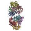
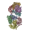
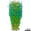
- PDB-5yys: Cryo-EM structure of L-fucokinase, GDP-fucose pyrophosphorylase (... -
+ Open data

- Basic information
| Entry | Database: PDB / ID: 5yys | |||||||||||||||||||||||||||||||||||||||||||||
|---|---|---|---|---|---|---|---|---|---|---|---|---|---|---|---|---|---|---|---|---|---|---|---|---|---|---|---|---|---|---|---|---|---|---|---|---|---|---|---|---|---|---|---|---|---|---|
| Title | Cryo-EM structure of L-fucokinase, GDP-fucose pyrophosphorylase (FKP)in Bacteroides fragilis | |||||||||||||||||||||||||||||||||||||||||||||
 Components Components | L-fucokinase, L-fucose-1-P guanylyltransferase | |||||||||||||||||||||||||||||||||||||||||||||
 Keywords Keywords | TRANSFERASE / bifunctional / kinase / ATP / GTP | |||||||||||||||||||||||||||||||||||||||||||||
| Function / homology |  Function and homology information Function and homology informationfucokinase / fucose-1-phosphate guanylyltransferase / fucose-1-phosphate guanylyltransferase activity / fucokinase activity / GDP-L-fucose salvage / ATP binding Similarity search - Function | |||||||||||||||||||||||||||||||||||||||||||||
| Biological species |  Bacteroides fragilis (bacteria) Bacteroides fragilis (bacteria) | |||||||||||||||||||||||||||||||||||||||||||||
| Method | ELECTRON MICROSCOPY / single particle reconstruction / cryo EM / Resolution: 4.2 Å | |||||||||||||||||||||||||||||||||||||||||||||
 Authors Authors | Wang, J. / Hu, H. / Liu, Y. / Zhou, Q. / Wu, P. / Yan, N. / Wang, H.W. / Wu, J.W. / Sun, L. | |||||||||||||||||||||||||||||||||||||||||||||
 Citation Citation | Journal: Protein Cell / Year: 2019 Title: Cryo-EM structure of L-fucokinase/GDP-fucose pyrophosphorylase (FKP) in Bacteroides fragilis. Authors: Ying Liu / Huifang Hu / Jia Wang / Qiang Zhou / Peng Wu / Nieng Yan / Hong-Wei Wang / Jia-Wei Wu / Linfeng Sun /   | |||||||||||||||||||||||||||||||||||||||||||||
| History |
|
- Structure visualization
| Movie |
Movie viewer |
|---|---|
| Structure viewer | Molecule: MolmilJmol/JSmol |
- Downloads & links
-Download
| PDBx/mmCIF format | 5yys.cif.gz | 552.4 KB | Display | PDBx/mmCIF format |
|---|---|---|---|---|
| PDB format | pdb5yys.ent.gz | 439.4 KB | Display | PDB format |
| PDBx/mmJSON format | 5yys.json.gz | Tree view | PDBx/mmJSON format | |
| Others |  Other downloads Other downloads |
-Validation report
| Arichive directory | https://data.pdbj.org/pub/pdb/validation_reports/yy/5yysftp://data.pdbj.org/pub/pdb/validation_reports/yy/5yys | HTTPS FTP |
|---|
-Related structure data
| Related structure data |  6859MC 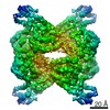 6860C  6861C M: map data used to model this data C: citing same article ( |
|---|---|
| Similar structure data |

-Links
 PDBj
PDBj- Assembly
| Deposited unit | 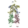
|
|---|---|
| 1 |
|
-Components
| #1: Protein | Mass: 105782.789 Da / Num. of mol.: 4 / Source method: isolated from a natural source / Source: (natural) Bacteroides fragilis (bacteria) / References: UniProt: Q58T34Has protein modification | N | |
|---|
-Experimental details
-Experiment
| Experiment | Method: ELECTRON MICROSCOPY |
|---|---|
| EM experiment | Aggregation state: PARTICLE / 3D reconstruction method: single particle reconstruction |
- Sample preparation
| Component | Name: FKP / Type: COMPLEX / Entity ID: all / Source: NATURAL |
|---|---|
| Source (natural) | Organism: Bacteroides fragilis (bacteria) |
| Buffer solution | pH: 8 |
| Specimen | Embedding applied: NO / Shadowing applied: NO / Staining applied: NO / Vitrification applied: YES |
| Specimen support | Grid material: COPPER / Grid mesh size: 400 divisions/in. / Grid type: Quantifoil R1.2/1.3 |
| Vitrification | Instrument: FEI VITROBOT MARK IV / Cryogen name: ETHANE / Humidity: 100 % |
- Electron microscopy imaging
| Experimental equipment |  Model: Titan Krios / Image courtesy: FEI Company |
|---|---|
| Microscopy | Model: FEI TITAN KRIOS |
| Electron gun | Electron source:  FIELD EMISSION GUN / Accelerating voltage: 300 kV / Illumination mode: FLOOD BEAM FIELD EMISSION GUN / Accelerating voltage: 300 kV / Illumination mode: FLOOD BEAM |
| Electron lens | Mode: BRIGHT FIELD |
| Image recording | Electron dose: 50 e/Å2 / Film or detector model: GATAN K2 SUMMIT (4k x 4k) |
- Processing
| Software | Name: PHENIX / Version: 1.12_2829: / Classification: refinement | ||||||||||||||||||||||||
|---|---|---|---|---|---|---|---|---|---|---|---|---|---|---|---|---|---|---|---|---|---|---|---|---|---|
| EM software | Name: PHENIX / Category: model refinement | ||||||||||||||||||||||||
| CTF correction | Type: PHASE FLIPPING AND AMPLITUDE CORRECTION | ||||||||||||||||||||||||
| 3D reconstruction | Resolution: 4.2 Å / Resolution method: FSC 0.143 CUT-OFF / Num. of particles: 96749 / Symmetry type: POINT | ||||||||||||||||||||||||
| Refine LS restraints |
|
