 Movie
Movie Controller

[English] 日本語
 Yorodumi
- PDB-6e47: Crystal Structure of the Murine Norovirus VP1 P domain in complex... -
+ Open data

- Basic information
| Entry | Database: PDB / ID: 6.0E+47 | |||||||||
|---|---|---|---|---|---|---|---|---|---|---|
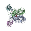
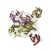
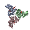
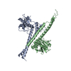
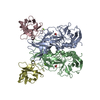
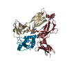
| Title | Crystal Structure of the Murine Norovirus VP1 P domain in complex with the CD300lf Receptor and Glycochenodeoxycholic Acid | |||||||||
 Components Components |
| |||||||||
 Keywords Keywords | VIRAL PROTEIN / Norovirus / Capsid protein / Protruding domain / bile acid / GCDCA / myeloid receptor / CMRF-35-like molecule-1 / CLM-1 / CLM1 / DIR2 / IREM1 / LMIR3 / PIGR3 / IgSF13 | |||||||||
| Function / homology |  Function and homology information Function and homology informationnegative regulation of MyD88-dependent toll-like receptor signaling pathway / interleukin-13-mediated signaling pathway / negative regulation of mast cell activation / interleukin-4 receptor binding / negative regulation of apoptotic cell clearance / positive regulation of interleukin-4-mediated signaling pathway / ceramide binding / positive regulation of apoptotic cell clearance / TRIF-dependent toll-like receptor signaling pathway / phosphatidylserine binding ...negative regulation of MyD88-dependent toll-like receptor signaling pathway / interleukin-13-mediated signaling pathway / negative regulation of mast cell activation / interleukin-4 receptor binding / negative regulation of apoptotic cell clearance / positive regulation of interleukin-4-mediated signaling pathway / ceramide binding / positive regulation of apoptotic cell clearance / TRIF-dependent toll-like receptor signaling pathway / phosphatidylserine binding / osteoclast differentiation / virion component / virus receptor activity / host cell cytoplasm / plasma membrane Similarity search - Function | |||||||||
| Biological species |  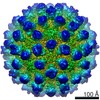 Murine norovirus 1 Murine norovirus 1 | |||||||||
| Method |  X-RAY DIFFRACTION / SYNCHROTRON / MOLECULAR REPLACEMENT / Resolution: 1.95 Å X-RAY DIFFRACTION / SYNCHROTRON / MOLECULAR REPLACEMENT / Resolution: 1.95 Å | |||||||||
 Authors Authors | Nelson, C.A. / Fremont, D.H. | |||||||||
| Funding support |  United States, 2items United States, 2items
| |||||||||
 Citation Citation | Journal: Proc Natl Acad Sci U S A / Year: 2018 Title: Structural basis for murine norovirus engagement of bile acids and the CD300lf receptor. Authors: Christopher A Nelson / Craig B Wilen / Ya-Nan Dai / Robert C Orchard / Arthur S Kim / Roderick A Stegeman / Leon L Hsieh / Thomas J Smith / Herbert W Virgin / Daved H Fremont / Abstract: Murine norovirus (MNoV) is closely related to human norovirus (HNoV), an infectious agent responsible for acute gastroenteritis worldwide. Here we report the X-ray crystal structure of the dimeric ...Murine norovirus (MNoV) is closely related to human norovirus (HNoV), an infectious agent responsible for acute gastroenteritis worldwide. Here we report the X-ray crystal structure of the dimeric MNoV VP1 protruding (P) domain in complex with its cellular receptor CD300lf. CD300lf binds the P domain with a 2:2 stoichiometry, engaging a cleft between the AB and DE loops of the P2 subdomain at a site that overlaps the epitopes of neutralizing antibodies. We also identify that bile acids are cofactors enhancing MNoV cell-binding and infectivity. Structures of CD300lf-P domain in complex with glycochenodeoxycholic acid (GCDCA) and lithocholic acid (LCA) reveal two bile acid binding sites at the P domain dimer interface distant from receptor binding sites. The structural determinants for receptor and bile acid binding are supported by numerous biophysical assays utilizing interface residue mutations. We find that the monomeric affinity of CD300lf for the P domain is low and is divalent cation dependent. We have also determined the crystal structure of CD300lf in complex with phosphocholine, revealing that MNoV engages its receptor in a manner mimicking host ligands including similar metal coordination. Docking of the cocomplex structures onto a cryo-EM-derived model of MNoV suggests that each virion can make multiple CD300lf engagements, and thus, infection may be driven by the avidity of cell surface clustered CD300lf. These studies identify multiple potential modulators of norovirus infection that may act to regulate the interaction between the viral capsid P domain and its cognate cellular receptor. | |||||||||
| History |
|
- Structure visualization
| Structure viewer | Molecule: MolmilJmol/JSmol |
|---|
- Downloads & links
-Download
| PDBx/mmCIF format | 6e47.cif.gz | 329.5 KB | Display | PDBx/mmCIF format |
|---|---|---|---|---|
| PDB format | pdb6e47.ent.gz | 268.8 KB | Display | PDB format |
| PDBx/mmJSON format | 6e47.json.gz | Tree view | PDBx/mmJSON format | |
| Others |  Other downloads Other downloads |
-Validation report
| Summary document | 6e47_validation.pdf.gz | 1022.9 KB | Display | wwPDB validaton report |
|---|---|---|---|---|
| Full document | 6e47_full_validation.pdf.gz | 1 MB | Display | |
| Data in XML | 6e47_validation.xml.gz | 37.1 KB | Display | |
| Data in CIF | 6e47_validation.cif.gz | 52.8 KB | Display | |
| Arichive directory | https://data.pdbj.org/pub/pdb/validation_reports/e4/6e47ftp://data.pdbj.org/pub/pdb/validation_reports/e4/6e47 | HTTPS FTP |
-Related structure data
| Related structure data | 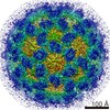 7564C 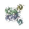 6c6qC 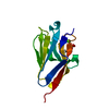 6c74C 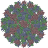 6crjC 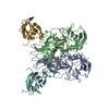 6e48C  3lq6S  5fflS S: Starting model for refinement C: citing same article ( |
|---|---|
| Similar structure data |


-Links
 PDBj
PDBj
- Assembly
| Deposited unit | 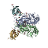
| ||||||||
|---|---|---|---|---|---|---|---|---|---|
| 1 |
| ||||||||
| Unit cell |
|
-Components
-Protein , 2 types, 4 molecules ABFG
| #1: Protein | Mass: 32906.094 Da / Num. of mol.: 2 / Fragment: VP1 Protruding domain (UNP residues 229-530) Source method: isolated from a genetically manipulated source Details: native / Source: (gene. exp.) Murine norovirus 1 / Plasmid: pET21a(+) / Production host:  #2: Protein | Mass: 12655.293 Da / Num. of mol.: 2 / Fragment: CD300lf ectodomain (UNP residues 20-131) Source method: isolated from a genetically manipulated source Details: refolded / Source: (gene. exp.) |
|---|
-Non-polymers , 4 types, 519 molecules 


| #3: Chemical | ChemComp-MG /  Mass: 24.305 Da / Num. of mol.: 6 / Source method: obtained synthetically / Formula: Mg Mass: 24.305 Da / Num. of mol.: 6 / Source method: obtained synthetically / Formula: Mg#4: Chemical | ChemComp-EDO /  Mass: 62.068 Da / Num. of mol.: 22 / Source method: obtained synthetically / Formula: C2H6O2 Mass: 62.068 Da / Num. of mol.: 22 / Source method: obtained synthetically / Formula: C2H6O2#5: Chemical |  Mass: 449.623 Da / Num. of mol.: 2 / Source method: obtained synthetically / Formula: C26H43NO5 / Feature type: SUBJECT OF INVESTIGATION / Comment: detergent*YM Mass: 449.623 Da / Num. of mol.: 2 / Source method: obtained synthetically / Formula: C26H43NO5 / Feature type: SUBJECT OF INVESTIGATION / Comment: detergent*YM#6: Water | ChemComp-HOH / | Mass: 18.015 Da / Num. of mol.: 489 / Source method: isolated from a natural source / Formula: H2O |
|---|
-Details
| Has protein modification | Y |
|---|
-Experimental details
-Experiment
| Experiment | Method: X-RAY DIFFRACTION / Number of used crystals: 1 |
|---|
- Sample preparation
| Crystal | Density Matthews: 2.54 Å3/Da / Density % sol: 51.49 % |
|---|---|
| Crystal grow | Temperature: 293 K / Method: vapor diffusion, hanging drop / pH: 8.7 Details: 0.1 M Tris, pH 8.7, 0.11 M magnesium chloride, 11% PEG10000, 100 uM sodium glycochenodeoxycholate |
-Data collection
| Diffraction | Mean temperature: 100 K |
|---|---|
| Diffraction source | Source: SYNCHROTRON / Site: ALS / Beamline: 4.2.2 / Wavelength: 1.00004 Å |
| Detector | Type: NOIR-1 / Detector: CMOS / Date: Oct 2, 2017 |
| Radiation | Monochromator: double crystal Si(111) / Protocol: SINGLE WAVELENGTH / Monochromatic (M) / Laue (L): M / Scattering type: x-ray |
| Radiation wavelength | Wavelength: 1.00004 Å / Relative weight: 1 |
| Reflection | Resolution: 1.95→60.7 Å / Num. obs: 67388 / % possible obs: 87.04 % / Redundancy: 5.8 % / CC1/2: 0.991 / Rmerge(I) obs: 0.2444 / Net I/σ(I): 6.48 |
| Reflection shell | Resolution: 1.95→2.02 Å / Num. unique obs: 1306 / CC1/2: 0.046 |
- Processing
| Software |
| |||||||||||||||||||||||||||||||||||||||||||||||||||||||||||||||||||||||||||||||||||||||||||||||||||||||||||||||||||||||||||||||||||||||||||||||||||||||||||||||||
|---|---|---|---|---|---|---|---|---|---|---|---|---|---|---|---|---|---|---|---|---|---|---|---|---|---|---|---|---|---|---|---|---|---|---|---|---|---|---|---|---|---|---|---|---|---|---|---|---|---|---|---|---|---|---|---|---|---|---|---|---|---|---|---|---|---|---|---|---|---|---|---|---|---|---|---|---|---|---|---|---|---|---|---|---|---|---|---|---|---|---|---|---|---|---|---|---|---|---|---|---|---|---|---|---|---|---|---|---|---|---|---|---|---|---|---|---|---|---|---|---|---|---|---|---|---|---|---|---|---|---|---|---|---|---|---|---|---|---|---|---|---|---|---|---|---|---|---|---|---|---|---|---|---|---|---|---|---|---|---|---|---|---|
| Refinement | Method to determine structure: MOLECULAR REPLACEMENT Starting model: PDB entries 3LQ6 & 5FFL Resolution: 1.95→60.7 Å / SU ML: 0.26 / Cross valid method: THROUGHOUT / σ(F): 1.34 / Phase error: 28.24
| |||||||||||||||||||||||||||||||||||||||||||||||||||||||||||||||||||||||||||||||||||||||||||||||||||||||||||||||||||||||||||||||||||||||||||||||||||||||||||||||||
| Solvent computation | Shrinkage radii: 0.9 Å / VDW probe radii: 1.11 Å | |||||||||||||||||||||||||||||||||||||||||||||||||||||||||||||||||||||||||||||||||||||||||||||||||||||||||||||||||||||||||||||||||||||||||||||||||||||||||||||||||
| Displacement parameters | Biso max: 133.54 Å2 / Biso mean: 38.196 Å2 / Biso min: 14.61 Å2 | |||||||||||||||||||||||||||||||||||||||||||||||||||||||||||||||||||||||||||||||||||||||||||||||||||||||||||||||||||||||||||||||||||||||||||||||||||||||||||||||||
| Refinement step | Cycle: final / Resolution: 1.95→60.7 Å
| |||||||||||||||||||||||||||||||||||||||||||||||||||||||||||||||||||||||||||||||||||||||||||||||||||||||||||||||||||||||||||||||||||||||||||||||||||||||||||||||||
| Refine LS restraints |
| |||||||||||||||||||||||||||||||||||||||||||||||||||||||||||||||||||||||||||||||||||||||||||||||||||||||||||||||||||||||||||||||||||||||||||||||||||||||||||||||||
| LS refinement shell | Refine-ID: X-RAY DIFFRACTION / Rfactor Rfree error: 0 / Total num. of bins used: 22
|
