 Movie
Movie Controller

[English] 日本語
 Yorodumi
- PDB-6crj: Mouse norovirus model using the crystal structure of MNV P domain... -
+ Open data

- Basic information
| Entry | Database: PDB / ID: 6crj | ||||||
|---|---|---|---|---|---|---|---|
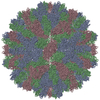
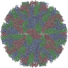
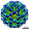
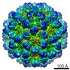
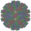
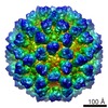
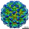
| Title | Mouse norovirus model using the crystal structure of MNV P domain and the Norwalkvirus shell domain | ||||||
 Components Components | Norwalk virus, MNV-1 capsid protein chimera | ||||||
 Keywords Keywords | VIRUS / norovirus / mouse | ||||||
| Function / homology |  Function and homology information Function and homology informationantigenic variation / T=3 icosahedral viral capsid / symbiont-mediated perturbation of host defense response / virion component / host cell cytoplasm / metal ion binding / identical protein binding Similarity search - Function | ||||||
| Biological species |  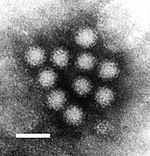 Norwalk virus Norwalk virus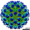 Murine norovirus 1 Murine norovirus 1 | ||||||
| Method | ELECTRON MICROSCOPY / single particle reconstruction / cryo EM / Resolution: 8 Å | ||||||
 Authors Authors | Smith, T.J. | ||||||
 Citation Citation | Journal: Proc Natl Acad Sci U S A / Year: 2018 Title: Structural basis for murine norovirus engagement of bile acids and the CD300lf receptor. Authors: Christopher A Nelson / Craig B Wilen / Ya-Nan Dai / Robert C Orchard / Arthur S Kim / Roderick A Stegeman / Leon L Hsieh / Thomas J Smith / Herbert W Virgin / Daved H Fremont /  Abstract: Murine norovirus (MNoV) is closely related to human norovirus (HNoV), an infectious agent responsible for acute gastroenteritis worldwide. Here we report the X-ray crystal structure of the dimeric ...Murine norovirus (MNoV) is closely related to human norovirus (HNoV), an infectious agent responsible for acute gastroenteritis worldwide. Here we report the X-ray crystal structure of the dimeric MNoV VP1 protruding (P) domain in complex with its cellular receptor CD300lf. CD300lf binds the P domain with a 2:2 stoichiometry, engaging a cleft between the AB and DE loops of the P2 subdomain at a site that overlaps the epitopes of neutralizing antibodies. We also identify that bile acids are cofactors enhancing MNoV cell-binding and infectivity. Structures of CD300lf-P domain in complex with glycochenodeoxycholic acid (GCDCA) and lithocholic acid (LCA) reveal two bile acid binding sites at the P domain dimer interface distant from receptor binding sites. The structural determinants for receptor and bile acid binding are supported by numerous biophysical assays utilizing interface residue mutations. We find that the monomeric affinity of CD300lf for the P domain is low and is divalent cation dependent. We have also determined the crystal structure of CD300lf in complex with phosphocholine, revealing that MNoV engages its receptor in a manner mimicking host ligands including similar metal coordination. Docking of the cocomplex structures onto a cryo-EM-derived model of MNoV suggests that each virion can make multiple CD300lf engagements, and thus, infection may be driven by the avidity of cell surface clustered CD300lf. These studies identify multiple potential modulators of norovirus infection that may act to regulate the interaction between the viral capsid P domain and its cognate cellular receptor. #1: Journal: J Virol / Year: 2010 Title: High-resolution cryo-electron microscopy structures of murine norovirus 1 and rabbit hemorrhagic disease virus reveal marked flexibility in the receptor binding domains. Authors: Umesh Katpally / Neil R Voss / Tommaso Cavazza / Stefan Taube / John R Rubin / Vivienne L Young / Jeanne Stuckey / Vernon K Ward / Herbert W Virgin / Christiane E Wobus / Thomas J Smith / Abstract: Our previous structural studies on intact, infectious murine norovirus 1 (MNV-1) virions demonstrated that the receptor binding protruding (P) domains are lifted off the inner shell of the virus. ...Our previous structural studies on intact, infectious murine norovirus 1 (MNV-1) virions demonstrated that the receptor binding protruding (P) domains are lifted off the inner shell of the virus. Here, the three-dimensional (3D) reconstructions of recombinant rabbit hemorrhagic disease virus (rRHDV) virus-like particles (VLPs) and intact MNV-1 were determined to approximately 8-A resolution. rRHDV also has a raised P domain, and therefore, this conformation is independent of infectivity and genus. The atomic structure of the MNV-1 P domain was used to interpret the MNV-1 reconstruction. Connections between the P and shell domains and between the floating P domains were modeled. This observed P-domain flexibility likely facilitates virus-host receptor interactions. | ||||||
| History |
|
- Structure visualization
| Movie |
Movie viewer |
|---|---|
| Structure viewer | Molecule: MolmilJmol/JSmol |
- Downloads & links
-Download
| PDBx/mmCIF format | 6crj.cif.gz | 293.7 KB | Display | PDBx/mmCIF format |
|---|---|---|---|---|
| PDB format | pdb6crj.ent.gz | 238.2 KB | Display | PDB format |
| PDBx/mmJSON format | 6crj.json.gz | Tree view | PDBx/mmJSON format | |
| Others |  Other downloads Other downloads |
-Validation report
| Arichive directory | https://data.pdbj.org/pub/pdb/validation_reports/cr/6crjftp://data.pdbj.org/pub/pdb/validation_reports/cr/6crj | HTTPS FTP |
|---|
-Related structure data
| Related structure data | 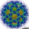 7564MC 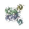 6c6qC 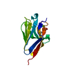 6c74C 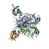 6e47C 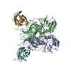 6e48C M: map data used to model this data C: citing same article ( |
|---|---|
| Similar structure data |

-Links
 PDBj
PDBj

- Assembly
| Deposited unit | 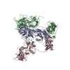
|
|---|---|
| 1 | x 60
|
| 2 |
|
| 3 | x 5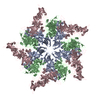
|
| 4 | x 6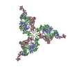
|
| 5 | 
|
| Symmetry | Point symmetry: (Schoenflies symbol: I (icosahedral)) |
-Components
| #1: Protein | Mass: 57127.523 Da / Num. of mol.: 3 Fragment: Norwalk shell domain (UNP residues 10-221), MNV-1 P domain (UNP residues 228-540) Source method: isolated from a genetically manipulated source Source: (gene. exp.) Norwalk virus, (gene. exp.) Murine norovirus 1Gene: ORF2 / Production host:  |
|---|
-Experimental details
-Experiment
| Experiment | Method: ELECTRON MICROSCOPY |
|---|---|
| EM experiment | Aggregation state: PARTICLE / 3D reconstruction method: single particle reconstruction |
- Sample preparation
| Component | Name: mouse norovirus model / Type: VIRUS Details: chimera of Norwalk virus shell domain and MNV P domain Entity ID: all / Source: RECOMBINANT |
|---|---|
| Source (natural) | Organism: Murine norovirus 1 |
| Source (recombinant) | Organism: |
| Details of virus | Empty: NO / Enveloped: NO / Isolate: STRAIN / Type: VIRION |
| Natural host | Organism: Mus |
| Buffer solution | pH: 7.2 / Details: PBS |
| Specimen | Embedding applied: NO / Shadowing applied: NO / Staining applied: NO / Vitrification applied: YES |
| Vitrification | Cryogen name: ETHANE |
- Electron microscopy imaging
| Experimental equipment |  Model: Tecnai F20 / Image courtesy: FEI Company |
|---|---|
| Microscopy | Model: FEI TECNAI F20 |
| Electron gun | Electron source:  FIELD EMISSION GUN / Accelerating voltage: 200 kV / Illumination mode: FLOOD BEAM FIELD EMISSION GUN / Accelerating voltage: 200 kV / Illumination mode: FLOOD BEAM |
| Electron lens | Mode: BRIGHT FIELD |
| Image recording | Electron dose: 10 e/Å2 / Film or detector model: FEI EAGLE (2k x 2k) |
- Processing
| CTF correction | Type: PHASE FLIPPING AND AMPLITUDE CORRECTION |
|---|---|
| 3D reconstruction | Resolution: 8 Å / Resolution method: FSC 0.5 CUT-OFF / Num. of particles: 20425 / Symmetry type: POINT |
| Atomic model building | Protocol: RIGID BODY FIT |
