 Movie
Movie Controller

[English] 日本語
 Yorodumi
- PDB-5h3w: The structure of the C-terminal of the fibronectin/fibrinogen-bin... -
+ Open data

- Basic information
| Entry | Database: PDB / ID: 5h3w | ||||||
|---|---|---|---|---|---|---|---|
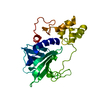
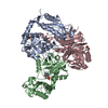
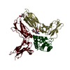
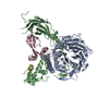
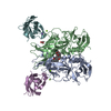
| Title | The structure of the C-terminal of the fibronectin/fibrinogen-binding protein from Streptococcus suis (FBPS) | ||||||
 Components Components | (Fibronectin/fibrinogen binding protein) x 2 | ||||||
 Keywords Keywords | CELL ADHESION / novel fold / fibronectin-binding property | ||||||
| Function / homology |  Function and homology information Function and homology informationRQC complex / ribosomal large subunit binding / rescue of stalled cytosolic ribosome / tRNA binding / cell adhesion / rRNA binding Similarity search - Function | ||||||
| Biological species |  Streptococcus suis (bacteria) Streptococcus suis (bacteria) | ||||||
| Method |  X-RAY DIFFRACTION / SYNCHROTRON / SAD / Resolution: 2.6 Å X-RAY DIFFRACTION / SYNCHROTRON / SAD / Resolution: 2.6 Å | ||||||
 Authors Authors | Musyoki, A.M. / Qi, J. / Gao, G.F. | ||||||
 Citation Citation | Journal: Proc. Natl. Acad. Sci. U.S.A. / Year: 2016 Title: Structural and functional analysis of an anchorless fibronectin-binding protein FBPS from Gram-positive bacterium Streptococcus suis Authors: Musyoki, A.M. / Shi, Z. / Xuan, C. / Lu, G. / Qi, J. / Gao, F. / Zheng, B. / Zhang, Q. / Li, Y. / Haywood, J. / Liu, C. / Yan, J. / Shi, Y. / Gao, G.F. | ||||||
| History |
|
- Structure visualization
| Structure viewer | Molecule: MolmilJmol/JSmol |
|---|
- Downloads & links
-Download
| PDBx/mmCIF format | 5h3w.cif.gz | 210.2 KB | Display | PDBx/mmCIF format |
|---|---|---|---|---|
| PDB format | pdb5h3w.ent.gz | 167.3 KB | Display | PDB format |
| PDBx/mmJSON format | 5h3w.json.gz | Tree view | PDBx/mmJSON format | |
| Others |  Other downloads Other downloads |
-Validation report
| Arichive directory | https://data.pdbj.org/pub/pdb/validation_reports/h3/5h3wftp://data.pdbj.org/pub/pdb/validation_reports/h3/5h3w | HTTPS FTP |
|---|
-Related structure data

-Links
 PDBj
PDBj- Assembly
| Deposited unit | 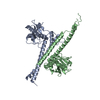
| ||||||||
|---|---|---|---|---|---|---|---|---|---|
| 1 |
| ||||||||
| Unit cell |
|
-Components
| #1: Protein | Mass: 32312.889 Da / Num. of mol.: 1 / Fragment: UNP residues 272-552 Source method: isolated from a genetically manipulated source Source: (gene. exp.) Streptococcus suis (bacteria) / Gene: fbps / Production host: |
|---|---|
| #2: Protein | Mass: 32470.084 Da / Num. of mol.: 1 / Fragment: UNP residues 271-552 / Mutation: L50I Source method: isolated from a genetically manipulated source Source: (gene. exp.) Streptococcus suis (bacteria) / Gene: fbps / Production host: |
| #3: Water | ChemComp-HOH /  Mass: 18.015 Da / Num. of mol.: 89 / Source method: isolated from a natural source / Formula: H2O Mass: 18.015 Da / Num. of mol.: 89 / Source method: isolated from a natural source / Formula: H2O |
-Experimental details
-Experiment
| Experiment | Method: X-RAY DIFFRACTION / Number of used crystals: 1 |
|---|
- Sample preparation
| Crystal | Density Matthews: 2.69 Å3/Da / Density % sol: 54.19 % |
|---|---|
| Crystal grow | Temperature: 291 K / Method: vapor diffusion, hanging drop Details: 0.1M HEPES (pH 7.5), 10%(w/v) PEG 4,000, 500mM NaCl |
-Data collection
| Diffraction | Mean temperature: 100 K |
|---|---|
| Diffraction source | Source: SYNCHROTRON / Site: SSRF  / Beamline: BL17U / Wavelength: 1 Å / Beamline: BL17U / Wavelength: 1 Å |
| Detector | Type: ADSC QUANTUM 315 / Detector: CCD / Date: Dec 1, 2009 |
| Radiation | Protocol: SINGLE WAVELENGTH / Monochromatic (M) / Laue (L): M / Scattering type: x-ray |
| Radiation wavelength | Wavelength: 1 Å / Relative weight: 1 |
| Reflection | Resolution: 2.6→50 Å / Num. obs: 21058 / % possible obs: 97.1 % / Redundancy: 6.5 % / Rmerge(I) obs: 0.115 / Net I/σ(I): 17.57 |
| Reflection shell | Resolution: 2.6→2.69 Å / Rmerge(I) obs: 0.338 |
- Processing
| Software |
| |||||||||||||||||||||||||||||||||||||||||||||||||||||||||||||||
|---|---|---|---|---|---|---|---|---|---|---|---|---|---|---|---|---|---|---|---|---|---|---|---|---|---|---|---|---|---|---|---|---|---|---|---|---|---|---|---|---|---|---|---|---|---|---|---|---|---|---|---|---|---|---|---|---|---|---|---|---|---|---|---|---|
| Refinement | Method to determine structure: SAD / Resolution: 2.6→38.613 Å / SU ML: 0.24 / Cross valid method: FREE R-VALUE / σ(F): 1.34 / Phase error: 27.72
| |||||||||||||||||||||||||||||||||||||||||||||||||||||||||||||||
| Solvent computation | Shrinkage radii: 0.9 Å / VDW probe radii: 1.11 Å | |||||||||||||||||||||||||||||||||||||||||||||||||||||||||||||||
| Refinement step | Cycle: LAST / Resolution: 2.6→38.613 Å
| |||||||||||||||||||||||||||||||||||||||||||||||||||||||||||||||
| Refine LS restraints |
| |||||||||||||||||||||||||||||||||||||||||||||||||||||||||||||||
| LS refinement shell |
| |||||||||||||||||||||||||||||||||||||||||||||||||||||||||||||||
| Refinement TLS params. | Method: refined / Origin x: 16.309 Å / Origin y: 19.6585 Å / Origin z: 13.004 Å
| |||||||||||||||||||||||||||||||||||||||||||||||||||||||||||||||
| Refinement TLS group | Selection details: all |
