 Movie
Movie Controller

[English] 日本語
 Yorodumi
- PDB-6d9r: The substrate-bound crystal structure of HPRT (hypoxanthine phosp... -
+ Open data

- Basic information
| Entry | Database: PDB / ID: 6d9r | ||||||||||||
|---|---|---|---|---|---|---|---|---|---|---|---|---|---|
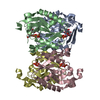
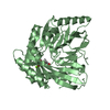
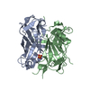
| Title | The substrate-bound crystal structure of HPRT (hypoxanthine phosphoribosyltransferase) | ||||||||||||
 Components Components | Hypoxanthine phosphoribosyltransferase | ||||||||||||
 Keywords Keywords | TRANSFERASE / HPRT / hypoxanthine phosphoribosyltransferase | ||||||||||||
| Function / homology |  Function and homology information Function and homology informationhypoxanthine phosphoribosyltransferase / guanine phosphoribosyltransferase activity / guanine salvage / hypoxanthine metabolic process / hypoxanthine phosphoribosyltransferase activity / GMP salvage / IMP salvage / purine ribonucleoside salvage / nucleotide binding / magnesium ion binding ...hypoxanthine phosphoribosyltransferase / guanine phosphoribosyltransferase activity / guanine salvage / hypoxanthine metabolic process / hypoxanthine phosphoribosyltransferase activity / GMP salvage / IMP salvage / purine ribonucleoside salvage / nucleotide binding / magnesium ion binding / metal ion binding / cytoplasm / cytosol Similarity search - Function | ||||||||||||
| Biological species |  | ||||||||||||
| Method |  X-RAY DIFFRACTION / SYNCHROTRON / MOLECULAR REPLACEMENT / Resolution: 1.64 Å X-RAY DIFFRACTION / SYNCHROTRON / MOLECULAR REPLACEMENT / Resolution: 1.64 Å | ||||||||||||
 Authors Authors | Satyshur, K.A. / Wolak, C. / Anderson, B. / Dubiel, K. / Keck, J.L. | ||||||||||||
| Funding support |  United States, 3items United States, 3items
| ||||||||||||
 Citation Citation | Journal: Elife / Year: 2019 Title: Evolution of (p)ppGpp-HPRT regulation through diversification of an allosteric oligomeric interaction. Authors: Anderson, B.W. / Liu, K. / Wolak, C. / Dubiel, K. / She, F. / Satyshur, K.A. / Keck, J.L. / Wang, J.D. | ||||||||||||
| History |
|
- Structure visualization
| Structure viewer | Molecule: MolmilJmol/JSmol |
|---|
- Downloads & links
-Download
| PDBx/mmCIF format | 6d9r.cif.gz | 164.2 KB | Display | PDBx/mmCIF format |
|---|---|---|---|---|
| PDB format | pdb6d9r.ent.gz | 131.8 KB | Display | PDB format |
| PDBx/mmJSON format | 6d9r.json.gz | Tree view | PDBx/mmJSON format | |
| Others |  Other downloads Other downloads |
-Validation report
| Arichive directory | https://data.pdbj.org/pub/pdb/validation_reports/d9/6d9rftp://data.pdbj.org/pub/pdb/validation_reports/d9/6d9r | HTTPS FTP |
|---|
-Related structure data
| Related structure data | 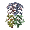 6d9qC  6d9sSC S: Starting model for refinement C: citing same article ( |
|---|---|
| Similar structure data |


-Links
 PDBj
PDBj
- Assembly
| Deposited unit | 
| |||||||||
|---|---|---|---|---|---|---|---|---|---|---|
| 1 |
| |||||||||
| Unit cell |
| |||||||||
| Components on special symmetry positions |
|
-Components
-Protein / Sugars , 2 types, 4 molecules AB
| #1: Protein | Mass: 20503.646 Da / Num. of mol.: 2 Source method: isolated from a genetically manipulated source Source: (gene. exp.) Gene: hpt, hprT, hpt1, tilS, BA_0063, A9486_05730, ABW01_27435, BASH2_00187, BVG01_28865, CN272_24955, CN488_12230, CN504_22375, COE56_22980, COJ30_24475, COK92_19380, COL95_25280, MCCC1A01412_27610 Plasmid: pLIC trPC-HA / Production host: References: UniProt: A0A1S0QLD4, UniProt: B9ZW32*PLUS, hypoxanthine phosphoribosyltransferase #3: Sugar |  Type: D-saccharide / Mass: 390.070 Da / Num. of mol.: 2 / Source method: obtained synthetically / Formula: C5H13O14P3 Type: D-saccharide / Mass: 390.070 Da / Num. of mol.: 2 / Source method: obtained synthetically / Formula: C5H13O14P3 |
|---|
-Non-polymers , 6 types, 342 molecules 


| #2: Chemical |  Mass: 150.138 Da / Num. of mol.: 2 / Source method: obtained synthetically / Formula: C6H6N4O Mass: 150.138 Da / Num. of mol.: 2 / Source method: obtained synthetically / Formula: C6H6N4O#4: Chemical | ChemComp-MG /  Mass: 24.305 Da / Num. of mol.: 4 / Source method: obtained synthetically / Formula: Mg Mass: 24.305 Da / Num. of mol.: 4 / Source method: obtained synthetically / Formula: Mg#5: Chemical | ChemComp-PEG /  Mass: 106.120 Da / Num. of mol.: 5 / Source method: obtained synthetically / Formula: C4H10O3 Mass: 106.120 Da / Num. of mol.: 5 / Source method: obtained synthetically / Formula: C4H10O3#6: Chemical |  Mass: 59.044 Da / Num. of mol.: 3 / Source method: obtained synthetically / Formula: C2H3O2 Mass: 59.044 Da / Num. of mol.: 3 / Source method: obtained synthetically / Formula: C2H3O2#7: Chemical | ChemComp-EDO /  Mass: 62.068 Da / Num. of mol.: 11 / Source method: obtained synthetically / Formula: C2H6O2 Mass: 62.068 Da / Num. of mol.: 11 / Source method: obtained synthetically / Formula: C2H6O2#8: Water | ChemComp-HOH / | Mass: 18.015 Da / Num. of mol.: 317 / Source method: isolated from a natural source / Formula: H2O |
|---|
-Experimental details
-Experiment
| Experiment | Method: X-RAY DIFFRACTION / Number of used crystals: 1 |
|---|
- Sample preparation
| Crystal | Density Matthews: 2.58 Å3/Da / Density % sol: 52.4 % |
|---|---|
| Crystal grow | Temperature: 293 K / Method: vapor diffusion, hanging drop Details: 0.2 M ammonium acetate, 0.1 M sodium acetate trihydrate, pH 4.6, 30% PEG4000 PH range: 4.6 - 8 |
-Data collection
| Diffraction | Mean temperature: 100 K |
|---|---|
| Diffraction source | Source: SYNCHROTRON / Site: APS / Beamline: 21-ID-F / Wavelength: 0.97872 Å |
| Detector | Type: RAYONIX MX-300 / Detector: CCD / Date: Mar 8, 2017 |
| Radiation | Monochromator: diamond(111) / Protocol: SINGLE WAVELENGTH / Monochromatic (M) / Laue (L): M / Scattering type: x-ray |
| Radiation wavelength | Wavelength: 0.97872 Å / Relative weight: 1 |
| Reflection | Resolution: 1.64→40.167 Å / Num. obs: 51750 / % possible obs: 99.8 % / Redundancy: 21.5 % / Biso Wilson estimate: 19.34 Å2 / Rpim(I) all: 0.029 / Rrim(I) all: 0.137 / Net I/σ(I): 31.09 |
| Reflection shell | Resolution: 1.64→1.67 Å / Redundancy: 14.4 % / Num. unique obs: 2548 / CC1/2: 0.81 / Rpim(I) all: 0.35 / Rrim(I) all: 1.376 / % possible all: 99 |
- Processing
| Software |
| |||||||||||||||||||||||||||||||||||||||||||||||||||||||||||||||||||||||||||||||||||||||||||||||||||||||||
|---|---|---|---|---|---|---|---|---|---|---|---|---|---|---|---|---|---|---|---|---|---|---|---|---|---|---|---|---|---|---|---|---|---|---|---|---|---|---|---|---|---|---|---|---|---|---|---|---|---|---|---|---|---|---|---|---|---|---|---|---|---|---|---|---|---|---|---|---|---|---|---|---|---|---|---|---|---|---|---|---|---|---|---|---|---|---|---|---|---|---|---|---|---|---|---|---|---|---|---|---|---|---|---|---|---|---|
| Refinement | Method to determine structure: MOLECULAR REPLACEMENT Starting model: PDB entry 6D9S Resolution: 1.64→37.19 Å / SU ML: 0.17 / Cross valid method: FREE R-VALUE / σ(F): 1.35 / Phase error: 20.44
| |||||||||||||||||||||||||||||||||||||||||||||||||||||||||||||||||||||||||||||||||||||||||||||||||||||||||
| Solvent computation | Shrinkage radii: 0.9 Å / VDW probe radii: 1.11 Å | |||||||||||||||||||||||||||||||||||||||||||||||||||||||||||||||||||||||||||||||||||||||||||||||||||||||||
| Refinement step | Cycle: LAST / Resolution: 1.64→37.19 Å
| |||||||||||||||||||||||||||||||||||||||||||||||||||||||||||||||||||||||||||||||||||||||||||||||||||||||||
| Refine LS restraints |
| |||||||||||||||||||||||||||||||||||||||||||||||||||||||||||||||||||||||||||||||||||||||||||||||||||||||||
| LS refinement shell |
|
