 Movie
Movie Controller

[English] 日本語
 Yorodumi
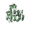
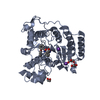
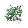
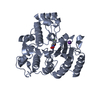
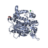
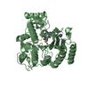
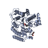
- PDB-4qa3: Crystal structure of T311M HDAC8 in complex with Trichostatin A (TSA) -
+ Open data

- Basic information
| Entry | Database: PDB / ID: 4qa3 | ||||||
|---|---|---|---|---|---|---|---|
| Title | Crystal structure of T311M HDAC8 in complex with Trichostatin A (TSA) | ||||||
 Components Components | Histone deacetylase 8 | ||||||
 Keywords Keywords | HYDROLASE / metalloenzyme / histone deacetylase / enzyme inhibitor complex / Cornelia de Lange Syndrome / arginase/deacetylase fold | ||||||
| Function / homology |  Function and homology information Function and homology informationhistone decrotonylase activity / histone deacetylase activity, hydrolytic mechanism / histone deacetylase / histone deacetylase activity / protein lysine deacetylase activity / Hydrolases; Acting on carbon-nitrogen bonds, other than peptide bonds; In linear amides / regulation of telomere maintenance / mitotic sister chromatid cohesion / Notch-HLH transcription pathway / nuclear chromosome ...histone decrotonylase activity / histone deacetylase activity, hydrolytic mechanism / histone deacetylase / histone deacetylase activity / protein lysine deacetylase activity / Hydrolases; Acting on carbon-nitrogen bonds, other than peptide bonds; In linear amides / regulation of telomere maintenance / mitotic sister chromatid cohesion / Notch-HLH transcription pathway / nuclear chromosome / histone deacetylase complex / negative regulation of protein ubiquitination / Hsp70 protein binding / Resolution of Sister Chromatid Cohesion / HDACs deacetylate histones / regulation of protein stability / Hsp90 protein binding / NOTCH1 Intracellular Domain Regulates Transcription / Constitutive Signaling by NOTCH1 PEST Domain Mutants / Constitutive Signaling by NOTCH1 HD+PEST Domain Mutants / Separation of Sister Chromatids / heterochromatin formation / chromatin organization / DNA-binding transcription factor binding / negative regulation of transcription by RNA polymerase II / nucleoplasm / metal ion binding / nucleus / cytoplasm Similarity search - Function | ||||||
| Biological species |  Homo sapiens (human) Homo sapiens (human) | ||||||
| Method |  X-RAY DIFFRACTION / SYNCHROTRON / MOLECULAR REPLACEMENT / Resolution: 2.876 Å X-RAY DIFFRACTION / SYNCHROTRON / MOLECULAR REPLACEMENT / Resolution: 2.876 Å | ||||||
 Authors Authors | Decroos, C. / Bowman, C.B. / Moser, J.-A.S. / Christianson, K.E. / Deardorff, M.A. / Christianson, D.W. | ||||||
 Citation Citation | Journal: Acs Chem.Biol. / Year: 2014 Title: Compromised Structure and Function of HDAC8 Mutants Identified in Cornelia de Lange Syndrome Spectrum Disorders. Authors: Decroos, C. / Bowman, C.M. / Moser, J.A. / Christianson, K.E. / Deardorff, M.A. / Christianson, D.W. | ||||||
| History |
|
- Structure visualization
| Structure viewer | Molecule: MolmilJmol/JSmol |
|---|
- Downloads & links
-Download
| PDBx/mmCIF format | 4qa3.cif.gz | 154.7 KB | Display | PDBx/mmCIF format |
|---|---|---|---|---|
| PDB format | pdb4qa3.ent.gz | 120.4 KB | Display | PDB format |
| PDBx/mmJSON format | 4qa3.json.gz | Tree view | PDBx/mmJSON format | |
| Others |  Other downloads Other downloads |
-Validation report
| Arichive directory | https://data.pdbj.org/pub/pdb/validation_reports/qa/4qa3ftp://data.pdbj.org/pub/pdb/validation_reports/qa/4qa3 | HTTPS FTP |
|---|
-Related structure data
| Related structure data |  4qa0C  4qa1C  4qa2C  4qa4C  4qa5C  4qa6C  4qa7C  3ewfS C: citing same article ( S: Starting model for refinement |
|---|---|
| Similar structure data |


-Links
 PDBj
PDBj


- Assembly
| Deposited unit | 
| ||||||||
|---|---|---|---|---|---|---|---|---|---|
| 1 | 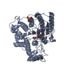
| ||||||||
| 2 | 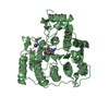
| ||||||||
| 3 |
| ||||||||
| Unit cell |
|
-Components
-Protein , 1 types, 2 molecules AB
| #1: Protein | Mass: 43262.086 Da / Num. of mol.: 2 / Mutation: T311M Source method: isolated from a genetically manipulated source Source: (gene. exp.) Homo sapiens (human) / Gene: HDAC8, HDACL1, CDA07 / Plasmid: pHD2-Xa-His / Production host:  |
|---|
-Non-polymers , 5 types, 43 molecules 


| #2: Chemical |  Mass: 302.368 Da / Num. of mol.: 2 / Source method: obtained synthetically / Formula: C17H22N2O3 Mass: 302.368 Da / Num. of mol.: 2 / Source method: obtained synthetically / Formula: C17H22N2O3#3: Chemical |  Mass: 65.409 Da / Num. of mol.: 2 / Source method: obtained synthetically / Formula: Zn Mass: 65.409 Da / Num. of mol.: 2 / Source method: obtained synthetically / Formula: Zn#4: Chemical | ChemComp-K /  Mass: 39.098 Da / Num. of mol.: 4 / Source method: obtained synthetically / Formula: K Mass: 39.098 Da / Num. of mol.: 4 / Source method: obtained synthetically / Formula: K#5: Chemical |  Mass: 92.094 Da / Num. of mol.: 2 / Source method: obtained synthetically / Formula: C3H8O3 Mass: 92.094 Da / Num. of mol.: 2 / Source method: obtained synthetically / Formula: C3H8O3#6: Water | ChemComp-HOH / | Mass: 18.015 Da / Num. of mol.: 33 / Source method: isolated from a natural source / Formula: H2O |
|---|
-Experimental details
-Experiment
| Experiment | Method: X-RAY DIFFRACTION / Number of used crystals: 1 |
|---|
- Sample preparation
| Crystal | Density Matthews: 2.31 Å3/Da / Density % sol: 46.64 % |
|---|---|
| Crystal grow | Temperature: 294 K / Method: vapor diffusion, sitting drop / pH: 8.3 Details: 0.1 M Bicine (pH = 8.5), 15% PEG 10000, VAPOR DIFFUSION, SITTING DROP, temperature 294K |
-Data collection
| Diffraction | Mean temperature: 100 K |
|---|---|
| Diffraction source | Source: SYNCHROTRON / Site: NSLS  / Beamline: X29A / Wavelength: 1.075 Å / Beamline: X29A / Wavelength: 1.075 Å |
| Detector | Type: ADSC QUANTUM 315r / Detector: CCD / Date: Apr 19, 2013 / Details: mirrors |
| Radiation | Monochromator: Si(111) / Protocol: SINGLE WAVELENGTH / Monochromatic (M) / Laue (L): M / Scattering type: x-ray |
| Radiation wavelength | Wavelength: 1.075 Å / Relative weight: 1 |
| Reflection | Resolution: 2.87→50 Å / Num. all: 18033 / Num. obs: 17775 / % possible obs: 98.6 % / Observed criterion σ(F): 0 / Observed criterion σ(I): -3 / Redundancy: 4.5 % / Biso Wilson estimate: 45.1 Å2 / Rmerge(I) obs: 0.12 / Net I/σ(I): 11.4 |
| Reflection shell | Resolution: 2.87→2.97 Å / Redundancy: 4.2 % / Rmerge(I) obs: 0.484 / Mean I/σ(I) obs: 3 / Num. unique all: 1647 / % possible all: 93.2 |
- Processing
| Software |
| ||||||||||||||||||||||||||||||||||||||||||||||||||||||||||||||||||||||||||||||||||||||||||||||||||
|---|---|---|---|---|---|---|---|---|---|---|---|---|---|---|---|---|---|---|---|---|---|---|---|---|---|---|---|---|---|---|---|---|---|---|---|---|---|---|---|---|---|---|---|---|---|---|---|---|---|---|---|---|---|---|---|---|---|---|---|---|---|---|---|---|---|---|---|---|---|---|---|---|---|---|---|---|---|---|---|---|---|---|---|---|---|---|---|---|---|---|---|---|---|---|---|---|---|---|---|
| Refinement | Method to determine structure: MOLECULAR REPLACEMENT Starting model: PDB ENTRY 3EWF Resolution: 2.876→46.896 Å / SU ML: 0.35 / Isotropic thermal model: isotropic / Cross valid method: THROUGHOUT / σ(F): 1.34 / Phase error: 24.52 / Stereochemistry target values: ML
| ||||||||||||||||||||||||||||||||||||||||||||||||||||||||||||||||||||||||||||||||||||||||||||||||||
| Solvent computation | Shrinkage radii: 0.9 Å / VDW probe radii: 1.11 Å / Solvent model: FLAT BULK SOLVENT MODEL | ||||||||||||||||||||||||||||||||||||||||||||||||||||||||||||||||||||||||||||||||||||||||||||||||||
| Displacement parameters | Biso mean: 44.24 Å2 | ||||||||||||||||||||||||||||||||||||||||||||||||||||||||||||||||||||||||||||||||||||||||||||||||||
| Refinement step | Cycle: LAST / Resolution: 2.876→46.896 Å
| ||||||||||||||||||||||||||||||||||||||||||||||||||||||||||||||||||||||||||||||||||||||||||||||||||
| Refine LS restraints |
| ||||||||||||||||||||||||||||||||||||||||||||||||||||||||||||||||||||||||||||||||||||||||||||||||||
| LS refinement shell |
|
