[English] 日本語
Yorodumi
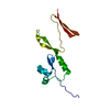
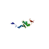
- PDB-6ckn: Crystal structure of an AF10 fragment -

+
Open data


ID or keywords:

Loading...

-
Basic information

Entry
Database: PDB / ID: 6ckn
TitleCrystal structure of an AF10 fragment
ComponentsProtein AF-10
KeywordsDNA BINDING PROTEIN / Structural Genomics / Structural Genomics Consortium / SGC
Function / homology
Function and homology information


nucleosome binding / histone binding / chromatin binding / regulation of transcription by RNA polymerase II / positive regulation of transcription by RNA polymerase II / protein-containing complex / DNA binding / nucleoplasm / metal ion binding / nucleus
Similarity search - Function
: / : / : / PHD-finger / PHD-zinc-finger like domain / Extended PHD (ePHD) domain / Extended PHD (ePHD) domain profile. / Zinc finger, PHD-type, conserved site / Zinc finger PHD-type signature. / Zinc finger PHD-type profile. ...: / : / : / PHD-finger / PHD-zinc-finger like domain / Extended PHD (ePHD) domain / Extended PHD (ePHD) domain profile. / Zinc finger, PHD-type, conserved site / Zinc finger PHD-type signature. / Zinc finger PHD-type profile. / Zinc finger, PHD-finger / Zinc finger, PHD-type / PHD zinc finger / Zinc finger, FYVE/PHD-type / Zinc finger, RING/FYVE/PHD-type
Similarity search - Domain/homology
Biological speciesHomo sapiens (human)
MethodX-RAY DIFFRACTION / SYNCHROTRON / SAD / Resolution: 2.49 Å
AuthorsQin, S. / Tempel, W. / Li, Y. / Bountra, C. / Arrowsmith, C.H. / Edwards, A.M. / Min, J. / Structural Genomics Consortium (SGC)
CitationJournal: Genes Dev. / Year: 2018
Title: Structural and functional analysis of the DOT1L-AF10 complex reveals mechanistic insights into MLL-AF10-associated leukemogenesis.
Authors: Zhang, H. / Zhou, B. / Qin, S. / Xu, J. / Harding, R. / Tempel, W. / Nayak, V. / Li, Y. / Loppnau, P. / Dou, Y. / Min, J.
History
DepositionFeb 28, 2018Deposition site: RCSB / Processing site: RCSB
Revision 1.0Mar 21, 2018Provider: repository / Type: Initial release
Revision 1.1Apr 18, 2018Group: Data collection / Database references / Category: citation / citation_author
Item: _citation.country / _citation.journal_abbrev ..._citation.country / _citation.journal_abbrev / _citation.journal_id_ASTM / _citation.journal_id_CSD / _citation.journal_id_ISSN / _citation.journal_volume / _citation.page_first / _citation.page_last / _citation.pdbx_database_id_DOI / _citation.pdbx_database_id_PubMed / _citation.title / _citation.year

-
Structure visualization

Structure viewerMolecule:
MolmilJmol/JSmol

Downloads & links

-
Assembly

Deposited unit
A: Protein AF-10
B: Protein AF-10


Theoretical massNumber of molelcules
Total (without water)17,5844
Polymers17,5842
Non-polymers02
Water0
1


  • Idetical with deposited unit
  • defined by author&software
TypeNameSymmetry operationNumber
identity operation1_555x,y,z1
Buried area1340 Å2
ΔGint-12 kcal/mol
Surface area9740 Å2
MethodPISA
Unit cell
Length a, b, c (Å)40.058, 40.058, 205.570
Angle α, β, γ (deg.)90.000, 90.000, 90.000
Int Tables number96
Space group name H-MP43212

-
Components

#1: Protein Protein AF-10 / ALL1-fused gene from chromosome 10 protein


Mass: 8791.813 Da / Num. of mol.: 2
Source method: isolated from a genetically manipulated source
Source: (gene. exp.) Homo sapiens (human) / Gene: MLLT10, AF10 / Production host: Escherichia coli (E. coli) / Strain (production host): BL21(DE3)-V2R-pRARE2 / References: UniProt: P55197
#2: Chemical ChemComp-UNX / UNKNOWN ATOM OR ION


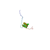
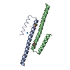
Num. of mol.: 2 / Source method: obtained synthetically

-
Experimental details

-
Experiment

ExperimentMethod: X-RAY DIFFRACTION / Number of used crystals: 1

-
Sample preparation

CrystalDensity Matthews: 2.36 Å3/Da / Density % sol: 47.83 %
Crystal growTemperature: 291 K / Method: vapor diffusion, sitting drop
Details: This crystal's crystallization record is incomplete. We attempted to reconstruct the condition through reference to records from related crystals: 20% PEG-3350, 0.2 M magnesium nitrate. Additive: 1,6-hexanediol

-
Data collection

DiffractionMean temperature: 100 K
Diffraction sourceSource: SYNCHROTRON / Site: APS / Beamline: 19-ID / Wavelength: 0.97918 Å
DetectorType: ADSC QUANTUM 315 / Detector: CCD / Date: Jun 20, 2013
RadiationProtocol: SINGLE WAVELENGTH / Monochromatic (M) / Laue (L): M / Scattering type: x-ray
Radiation wavelengthWavelength: 0.97918 Å / Relative weight: 1
ReflectionResolution: 2.49→41.11 Å / Num. obs: 6558 / % possible obs: 99.9 % / Redundancy: 13.1 % / Biso Wilson estimate: 66.04 Å2 / CC1/2: 0.999 / Rmerge(I) obs: 0.087 / Rpim(I) all: 0.025 / Rrim(I) all: 0.091 / Net I/σ(I): 20.6 / Num. measured all: 85873 / Scaling rejects: 2
Reflection shellResolution: 2.49→2.59 Å / Redundancy: 13.7 % / Rmerge(I) obs: 1.272 / Num. unique obs: 696 / CC1/2: 0.963 / Rpim(I) all: 0.354 / Rrim(I) all: 1.322 / % possible all: 99.8

-
Processing

Software
NameVersionClassification
BUSTER2.10.2refinement
Aimless0.5.32data scaling
PDB_EXTRACT3.22data extraction
XDSdata reduction
PHASERphasing
SHELXDphasing
PARROTphasing
BUCCANEERmodel building
RefinementMethod to determine structure: SAD / Resolution: 2.49→39.32 Å / Cor.coef. Fo:Fc: 0.888 / Cor.coef. Fo:Fc free: 0.901 / Rfactor Rfree error: 0 / SU R Cruickshank DPI: 0.377 / Cross valid method: THROUGHOUT / σ(F): 0 / SU R Blow DPI: 0.407 / SU Rfree Blow DPI: 0.257 / SU Rfree Cruickshank DPI: 0.253
Details: Se-Met SAD phasing with shelx/phaser/parrot. Autotracing with BUCCANEER. Refinement with REFMAC and autobuster.
RfactorNum. reflection% reflection
Rfree0.262 652 10.05 %
Rwork0.247 --
obs0.249 6488 99.9 %
Displacement parametersBiso max: 150.5 Å2 / Biso mean: 79.69 Å2 / Biso min: 55.09 Å2
Baniso -1Baniso -2Baniso -3
1--18.15 Å20 Å20 Å2
2---18.15 Å20 Å2
3---36.2999 Å2
Refine analyzeLuzzati coordinate error obs: 0.44 Å
Refinement stepCycle: final / Resolution: 2.49→39.32 Å
ProteinNucleic acidLigandSolventTotal
Num. atoms973 0 2 0 975
Biso mean--61.64 --
Num. residues----132
Refine LS restraints
Refine-IDTypeNumberRestraint functionWeightDev ideal
X-RAY DIFFRACTIONt_dihedral_angle_d353SINUSOIDAL2
X-RAY DIFFRACTIONt_trig_c_planes31HARMONIC2
X-RAY DIFFRACTIONt_gen_planes140HARMONIC5
X-RAY DIFFRACTIONt_it980HARMONIC20
X-RAY DIFFRACTIONt_nbd
X-RAY DIFFRACTIONt_improper_torsion
X-RAY DIFFRACTIONt_pseud_angle
X-RAY DIFFRACTIONt_chiral_improper_torsion133SEMIHARMONIC5
X-RAY DIFFRACTIONt_sum_occupancies
X-RAY DIFFRACTIONt_utility_distance
X-RAY DIFFRACTIONt_utility_angle
X-RAY DIFFRACTIONt_utility_torsion
X-RAY DIFFRACTIONt_ideal_dist_contact1163SEMIHARMONIC4
X-RAY DIFFRACTIONt_bond_d980HARMONIC20.01
X-RAY DIFFRACTIONt_angle_deg1323HARMONIC21.1
X-RAY DIFFRACTIONt_omega_torsion2.3
X-RAY DIFFRACTIONt_other_torsion14.95
LS refinement shellResolution: 2.49→2.78 Å / Total num. of bins used: 5
RfactorNum. reflection% reflection
Rfree0.28 183 10.45 %
Rwork0.278 1569 -
all-1752 -
obs--99.89 %
Refinement TLS params.

Method: refined / Refine-ID: X-RAY DIFFRACTION

IDL112)L122)L132)L222)L232)L332)S11 (Å °)S12 (Å °)S13 (Å °)S21 (Å °)S22 (Å °)S23 (Å °)S31 (Å °)S32 (Å °)S33 (Å °)T112)T122)T132)T222)T232)T332)Origin x (Å)Origin y (Å)Origin z (Å)
12.11180.5835-2.50720-0.65082.91720.0368-0.05140.17860.24530.0407-0.073-0.1833-0.5199-0.07750.0613-0.1598-0.05530.0031-0.0002-0.1825-31.559633.0791109.74
21.02811.7485-0.26881.8838-2.85342.97160.02170.3008-0.0916-0.12160.0511-0.0047-0.5056-0.1979-0.07270.0863-0.095-0.0073-0.0924-0.0493-0.2007-36.704125.478395.1806
Refinement TLS group
IDRefine-IDRefine TLS-IDSelection detailsAuth asym-IDAuth seq-ID
1X-RAY DIFFRACTION1{ A|* }A730 - 795
2X-RAY DIFFRACTION2{ B|* }B730 - 795

+
About Yorodumi

-
News

-
Feb 9, 2022. New format data for meta-information of EMDB entries

New format data for meta-information of EMDB entries

  • Version 3 of the EMDB header file is now the official format.
  • The previous official version 1.9 will be removed from the archive.

Related info.:EMDB header

External links:wwPDB to switch to version 3 of the EMDB data model

-
Aug 12, 2020. Covid-19 info

Covid-19 info

URL: https://pdbj.org/emnavi/covid19.php

New page: Covid-19 featured information page in EM Navigator.

Related info.:Covid-19 info / Mar 5, 2020. Novel coronavirus structure data

+
Mar 5, 2020. Novel coronavirus structure data

Novel coronavirus structure data

Related info.:Yorodumi Speices / Aug 12, 2020. Covid-19 info

External links:COVID-19 featured content - PDBj / Molecule of the Month (242):Coronavirus Proteases

+
Jan 31, 2019. EMDB accession codes are about to change! (news from PDBe EMDB page)

EMDB accession codes are about to change! (news from PDBe EMDB page)

  • The allocation of 4 digits for EMDB accession codes will soon come to an end. Whilst these codes will remain in use, new EMDB accession codes will include an additional digit and will expand incrementally as the available range of codes is exhausted. The current 4-digit format prefixed with “EMD-” (i.e. EMD-XXXX) will advance to a 5-digit format (i.e. EMD-XXXXX), and so on. It is currently estimated that the 4-digit codes will be depleted around Spring 2019, at which point the 5-digit format will come into force.
  • The EM Navigator/Yorodumi systems omit the EMD- prefix.

Related info.:Q: What is EMD? / ID/Accession-code notation in Yorodumi/EM Navigator

External links:EMDB Accession Codes are Changing Soon! / Contact to PDBj

+
Jul 12, 2017. Major update of PDB

Major update of PDB

  • wwPDB released updated PDB data conforming to the new PDBx/mmCIF dictionary.
  • This is a major update changing the version number from 4 to 5, and with Remediation, in which all the entries are updated.
  • In this update, many items about electron microscopy experimental information are reorganized (e.g. em_software).
  • Now, EM Navigator and Yorodumi are based on the updated data.

External links:wwPDB Remediation / Enriched Model Files Conforming to OneDep Data Standards Now Available in the PDB FTP Archive

-
Yorodumi

Thousand views of thousand structures

  • Yorodumi is a browser for structure data from EMDB, PDB, SASBDB, etc.
  • This page is also the successor to EM Navigator detail page, and also detail information page/front-end page for Omokage search.
  • The word "yorodu" (or yorozu) is an old Japanese word meaning "ten thousand". "mi" (miru) is to see.

Related info.:EMDB / PDB / SASBDB / Comparison of 3 databanks / Yorodumi Search / Aug 31, 2016. New EM Navigator & Yorodumi / Yorodumi Papers / Jmol/JSmol / Function and homology information / Changes in new EM Navigator and Yorodumi

Read more