 Movie
Movie Controller

[English] 日本語
 Yorodumi
- PDB-6c85: Crystal structure of aspartate semialdehyde dehydrogenase from Bl... -
+ Open data

- Basic information
| Entry | Database: PDB / ID: 6c85 | ||||||
|---|---|---|---|---|---|---|---|
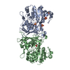
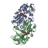
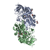
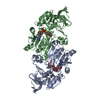
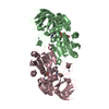
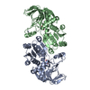
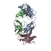
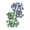
| Title | Crystal structure of aspartate semialdehyde dehydrogenase from Blastomyces dermatitidis with p-benzoquinone | ||||||
 Components Components | Aspartate-semialdehyde dehydrogenase | ||||||
 Keywords Keywords | OXIDOREDUCTASE / Rossman fold / Antifungal inhibitor complex | ||||||
| Function / homology |  Function and homology information Function and homology informationaspartate-semialdehyde dehydrogenase / aspartate-semialdehyde dehydrogenase activity / L-threonine biosynthetic process / : / NAD binding / NADP binding / protein dimerization activity Similarity search - Function | ||||||
| Biological species |  Blastomyces gilchristii (fungus) Blastomyces gilchristii (fungus) | ||||||
| Method |  X-RAY DIFFRACTION / SYNCHROTRON / MOLECULAR REPLACEMENT / Resolution: 2.4 Å X-RAY DIFFRACTION / SYNCHROTRON / MOLECULAR REPLACEMENT / Resolution: 2.4 Å | ||||||
 Authors Authors | Dahal, G.P. / Viola, R.E. | ||||||
| Funding support |  United States, 1items United States, 1items
| ||||||
 Citation Citation | Journal: Biochem.Biophys.Res.Commun. / Year: 2018 Title: Structural insights into inhibitor binding to a fungal ortholog of aspartate semialdehyde dehydrogenase. Authors: Dahal, G.P. / Viola, R.E. | ||||||
| History |
|
- Structure visualization
| Structure viewer | Molecule: MolmilJmol/JSmol |
|---|
- Downloads & links
-Download
| PDBx/mmCIF format | 6c85.cif.gz | 144.4 KB | Display | PDBx/mmCIF format |
|---|---|---|---|---|
| PDB format | pdb6c85.ent.gz | 113.7 KB | Display | PDB format |
| PDBx/mmJSON format | 6c85.json.gz | Tree view | PDBx/mmJSON format | |
| Others |  Other downloads Other downloads |
-Validation report
| Arichive directory | https://data.pdbj.org/pub/pdb/validation_reports/c8/6c85ftp://data.pdbj.org/pub/pdb/validation_reports/c8/6c85 | HTTPS FTP |
|---|
-Related structure data
| Related structure data |  6c8wC  3hskS S: Starting model for refinement C: citing same article ( |
|---|---|
| Similar structure data |


-Links
 PDBj
PDBj- Assembly
| Deposited unit | 
| ||||||||
|---|---|---|---|---|---|---|---|---|---|
| 1 |
| ||||||||
| Unit cell |
| ||||||||
| Components on special symmetry positions |
|
-Components
| #1: Protein | Mass: 40152.977 Da / Num. of mol.: 2 Source method: isolated from a genetically manipulated source Source: (gene. exp.) Blastomyces gilchristii (fungus) / Strain: SLH14081 / Gene: BDCG_01946 / Plasmid: pET-28a(+) / Cell line (production host): BL21 (DE3) / Production host:  #2: Chemical |   Mass: 108.095 Da / Num. of mol.: 2 / Source method: obtained synthetically / Formula: C6H4O2 Mass: 108.095 Da / Num. of mol.: 2 / Source method: obtained synthetically / Formula: C6H4O2#3: Water | ChemComp-HOH / |  Mass: 18.015 Da / Num. of mol.: 102 / Source method: isolated from a natural source / Formula: H2O Mass: 18.015 Da / Num. of mol.: 102 / Source method: isolated from a natural source / Formula: H2O |
|---|
-Experimental details
-Experiment
| Experiment | Method: X-RAY DIFFRACTION / Number of used crystals: 1 |
|---|
- Sample preparation
| Crystal | Density Matthews: 2.32 Å3/Da / Density % sol: 47 % / Description: long rod-shaped |
|---|---|
| Crystal grow | Temperature: 293 K / Method: vapor diffusion, hanging drop / pH: 7 Details: 0.2M Ammonium Citrate Tribasic pH 7.0, 18 % w/v PEG 3350 PH range: 6.5-8.0 |
-Data collection
| Diffraction | Mean temperature: 100 K |
|---|---|
| Diffraction source | Source: SYNCHROTRON / Site: APS / Beamline: 23-ID-B / Wavelength: 1.0332 Å |
| Detector | Type: DECTRIS EIGER X 16M / Detector: PIXEL / Date: Apr 24, 2017 |
| Radiation | Protocol: SINGLE WAVELENGTH / Monochromatic (M) / Laue (L): M / Scattering type: x-ray |
| Radiation wavelength | Wavelength: 1.0332 Å / Relative weight: 1 |
| Reflection | Resolution: 2.4→85.8 Å / Num. obs: 59089 / % possible obs: 100 % / Redundancy: 9.7 % / Biso Wilson estimate: 52.2 Å2 / CC1/2: 0.999 / Rmerge(I) obs: 0.049 / Net I/σ(I): 15.7 |
| Reflection shell | Resolution: 2.4→2.47 Å / Redundancy: 9.7 % / Mean I/σ(I) obs: 2.96 / Num. unique obs: 4511 / CC1/2: 0.953 / % possible all: 100 |
- Processing
| Software |
| ||||||||||||||||||||||||
|---|---|---|---|---|---|---|---|---|---|---|---|---|---|---|---|---|---|---|---|---|---|---|---|---|---|
| Refinement | Method to determine structure: MOLECULAR REPLACEMENT Starting model: 3hsk Resolution: 2.4→71.417 Å / SU ML: 0.32 / Cross valid method: THROUGHOUT / σ(F): 1.34 / Phase error: 28.58 / Stereochemistry target values: ML
| ||||||||||||||||||||||||
| Solvent computation | Shrinkage radii: 0.9 Å / VDW probe radii: 1.11 Å / Solvent model: FLAT BULK SOLVENT MODEL | ||||||||||||||||||||||||
| Displacement parameters | Biso max: 150.8 Å2 / Biso mean: 64.6548 Å2 / Biso min: 34.16 Å2 | ||||||||||||||||||||||||
| Refinement step | Cycle: final / Resolution: 2.4→71.417 Å
|
