Mass: 18.015 Da / Num. of mol.: 17 / Source method: isolated from a natural source / Formula: H2O
Compound details
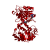
ENGINEERED RESIDUE IN CHAIN A, ASP 270 TO ASN ENGINEERED RESIDUE IN CHAIN B, ASP 270 TO ASN
Sequence details
EXPRESSED WITH A C-TERMINAL 6XHIS TAG.
-
Experimental details
-
Experiment
Experiment
Method: X-RAY DIFFRACTION / Number of used crystals: 1
-
Sample preparation
Crystal
Density Matthews: 3 Å3/Da / Density % sol: 58.62 % / Description: NONE
Crystal grow
pH: 7 Details: 0.1M BIS TRIS PROPANE PH 8.2, 20% PEG 3350 AND 0.2M SODIUM ACETATE. CO CRYSTALLIZED WITH 10MM NADH WHICH WAS ADDED TO THE PROTEIN SOLUTION
Monochromator: DIAMOND (111), GE(220) / Protocol: SINGLE WAVELENGTH / Monochromatic (M) / Laue (L): M / Scattering type: x-ray
Radiation wavelength
Wavelength: 0.934 Å / Relative weight: 1
Reflection
Resolution: 2.8→40 Å / Num. obs: 22947 / % possible obs: 99 % / Observed criterion σ(I): 4.1 / Redundancy: 4.4 % / Rmerge(I) obs: 0.08 / Net I/σ(I): 12.9
Reflection shell
Resolution: 2.8→2.95 Å / Redundancy: 4.5 % / Rmerge(I) obs: 0.4 / Mean I/σ(I) obs: 4.1 / % possible all: 99
-
Processing
Software
Name
Version
Classification
REFMAC
5.2.0019
refinement
MOSFLM
datareduction
SCALA
datascaling
MOLREP
phasing
Refinement
Method to determine structure: MOLECULAR REPLACEMENT / Resolution: 2.8→40 Å / Cor.coef. Fo:Fc: 0.934 / Cor.coef. Fo:Fc free: 0.904 / SU B: 25.753 / SU ML: 0.248 / Cross valid method: THROUGHOUT / ESU R Free: 0.349 / Stereochemistry target values: MAXIMUM LIKELIHOOD / Details: HYDROGENS HAVE BEEN ADDED IN THE RIDING POSITIONS
Rfactor
Num. reflection
% reflection
Selection details
Rfree
0.242
1162
5.1 %
RANDOM
Rwork
0.2
-
-
-
obs
0.202
21753
98.9 %
-
Solvent computation
Ion probe radii: 0.8 Å / Shrinkage radii: 0.8 Å / VDW probe radii: 1.4 Å / Solvent model: BABINET MODEL WITH MASK
 Movie
Movie Controller

 Yorodumi
 Open data
 Basic information
 Components
 Keywords
 Function and homology information
 MYCOBACTERIUM TUBERCULOSIS (bacteria)
 X-RAY DIFFRACTION /
  Authors
 Citation
 Structure visualization
 Downloads & links
 Other downloads


 PDBj
PDBj

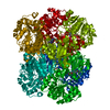
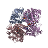
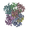
 Assembly


 Mass: 665.441 Da / Num. of mol.: 2 / Source method: obtained synthetically / Formula: C21H29N7O14P2
 Mass: 18.015 Da / Num. of mol.: 17 / Source method: isolated from a natural source / Formula: H2O
 Sample preparation
 / Beamline: ID14-1 / Wavelength: 0.934
  Processing