 Movie
Movie Controller

[English] 日本語
 Yorodumi
- PDB-2vhy: Crystal structure of apo L-alanine dehydrogenase from Mycobacteri... -
+ Open data

- Basic information
| Entry | Database: PDB / ID: 2vhy | ||||||
|---|---|---|---|---|---|---|---|
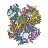
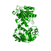
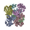
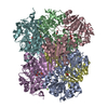
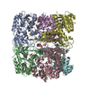
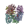
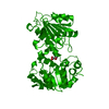
| Title | Crystal structure of apo L-alanine dehydrogenase from Mycobacterium tuberculosis | ||||||
 Components Components | ALANINE DEHYDROGENASE | ||||||
 Keywords Keywords | OXIDOREDUCTASE / NAD / SECRETED | ||||||
| Function / homology |  Function and homology information Function and homology information: / alanine dehydrogenase / L-alanine dehydrogenase (NAD+) activity / L-alanine catabolic process / cell wall / peptidoglycan-based cell wall / response to hypoxia / nucleotide binding / extracellular region / metal ion binding ...: / alanine dehydrogenase / L-alanine dehydrogenase (NAD+) activity / L-alanine catabolic process / cell wall / peptidoglycan-based cell wall / response to hypoxia / nucleotide binding / extracellular region / metal ion binding / plasma membrane / cytosol Similarity search - Function | ||||||
| Biological species |   MYCOBACTERIUM TUBERCULOSIS (bacteria) MYCOBACTERIUM TUBERCULOSIS (bacteria) | ||||||
| Method |  X-RAY DIFFRACTION / SYNCHROTRON / MOLECULAR REPLACEMENT / Resolution: 2.3 Å X-RAY DIFFRACTION / SYNCHROTRON / MOLECULAR REPLACEMENT / Resolution: 2.3 Å | ||||||
 Authors Authors | Agren, D. / Schneider, G. | ||||||
 Citation Citation | Journal: J.Mol.Biol. / Year: 2008 Title: Three-Dimensional Structures of Apo- and Holo-L-Alanine Dehydrogenase from Mycobacterium Tuberculosis Reveal Conformational Changes Upon Coenzyme Binding. Authors: Agren, D. / Stehr, M. / Berthold, C.L. / Kapoor, S. / Oehlmann, W. / Singh, M. / Schneider, G. | ||||||
| History |
|
- Structure visualization
| Structure viewer | Molecule: MolmilJmol/JSmol |
|---|
- Downloads & links
-Download
| PDBx/mmCIF format | 2vhy.cif.gz | 131.1 KB | Display | PDBx/mmCIF format |
|---|---|---|---|---|
| PDB format | pdb2vhy.ent.gz | 102.3 KB | Display | PDB format |
| PDBx/mmJSON format | 2vhy.json.gz | Tree view | PDBx/mmJSON format | |
| Others |  Other downloads Other downloads |
-Validation report
| Arichive directory | https://data.pdbj.org/pub/pdb/validation_reports/vh/2vhyftp://data.pdbj.org/pub/pdb/validation_reports/vh/2vhy | HTTPS FTP |
|---|
-Related structure data
| Related structure data |  2vhvC  2vhwC  2vhxC  2vhzC  1pjcS C: citing same article ( S: Starting model for refinement |
|---|---|
| Similar structure data |

-Links
 PDBj
PDBj- Assembly
| Deposited unit | 
| ||||||||
|---|---|---|---|---|---|---|---|---|---|
| 1 | 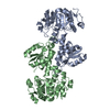
| ||||||||
| Unit cell |
| ||||||||
| Components on special symmetry positions |
| ||||||||
| Noncrystallographic symmetry (NCS) | NCS oper: (Code: given Matrix: (-0.97754, 0.21005, 0.01697), Vector: |
-Components
| #1: Protein | Mass: 39581.961 Da / Num. of mol.: 2 Source method: isolated from a genetically manipulated source Source: (gene. exp.) MYCOBACTERIUM TUBERCULOSIS (bacteria) / Plasmid: PET26 / Production host: References: UniProt: P30234, UniProt: P9WQB1*PLUS, alanine dehydrogenase #2: Water | ChemComp-HOH / |  Mass: 18.015 Da / Num. of mol.: 48 / Source method: isolated from a natural source / Formula: H2O Mass: 18.015 Da / Num. of mol.: 48 / Source method: isolated from a natural source / Formula: H2OSequence details | EXPRESSED WITH A C-TERMINAL 6XHIS TAG. | |
|---|
-Experimental details
-Experiment
| Experiment | Method: X-RAY DIFFRACTION / Number of used crystals: 1 |
|---|
- Sample preparation
| Crystal | Density Matthews: 4.31 Å3/Da / Density % sol: 71.24 % / Description: NONE |
|---|---|
| Crystal grow | Details: 0.4M AMMONIUM BICARBONATE. |
-Data collection
| Diffraction | Mean temperature: 100 K |
|---|---|
| Diffraction source | Source: SYNCHROTRON / Site: MAX II  / Beamline: I911-3 / Wavelength: 1 / Beamline: I911-3 / Wavelength: 1 |
| Detector | Type: MARRESEARCH / Detector: CCD / Date: Oct 6, 2006 / Details: RH-COATED TOROIDAL SI MIRROR |
| Radiation | Protocol: SINGLE WAVELENGTH / Monochromatic (M) / Laue (L): M / Scattering type: x-ray |
| Radiation wavelength | Wavelength: 1 Å / Relative weight: 1 |
| Reflection | Resolution: 2.3→45 Å / Num. obs: 47413 / % possible obs: 97 % / Observed criterion σ(I): 2.9 / Redundancy: 7.3 % / Rmerge(I) obs: 0.08 / Net I/σ(I): 21 |
| Reflection shell | Resolution: 2.34→2.47 Å / Redundancy: 6.1 % / Rmerge(I) obs: 0.49 / Mean I/σ(I) obs: 2.9 / % possible all: 79 |
- Processing
| Software |
| ||||||||||||||||||||||||||||||||||||||||||||||||||||||||||||
|---|---|---|---|---|---|---|---|---|---|---|---|---|---|---|---|---|---|---|---|---|---|---|---|---|---|---|---|---|---|---|---|---|---|---|---|---|---|---|---|---|---|---|---|---|---|---|---|---|---|---|---|---|---|---|---|---|---|---|---|---|---|
| Refinement | Method to determine structure: MOLECULAR REPLACEMENT Starting model: PDB ENTRY 1PJC Resolution: 2.3→45 Å / Data cutoff high absF: 10000 / Cross valid method: THROUGHOUT / σ(F): 0 Stereochemistry target values: LEAST SQUARES RESIDUAL FOR HEMIHEDRAL TWINNING Details: THE PROTOCOLL FOR HEMIHEDRALLY TWINNED DATA WAS USED. THE R-VALUES ARE FOR TWINNED DATA. RESIDUES A239-A258, A269-A294, A297-A301, B242-B255, B268- B293 AND B298-B301 ARE REMOVED FROM THE ...Details: THE PROTOCOLL FOR HEMIHEDRALLY TWINNED DATA WAS USED. THE R-VALUES ARE FOR TWINNED DATA. RESIDUES A239-A258, A269-A294, A297-A301, B242-B255, B268- B293 AND B298-B301 ARE REMOVED FROM THE MODEL SINCE THERE IS NO DENSITY FOR THESE RESIDUES.
| ||||||||||||||||||||||||||||||||||||||||||||||||||||||||||||
| Solvent computation | Bsol: 50.2348 Å2 / ksol: 0.372117 e/Å3 | ||||||||||||||||||||||||||||||||||||||||||||||||||||||||||||
| Displacement parameters |
| ||||||||||||||||||||||||||||||||||||||||||||||||||||||||||||
| Refinement step | Cycle: LAST / Resolution: 2.3→45 Å
| ||||||||||||||||||||||||||||||||||||||||||||||||||||||||||||
| Refine LS restraints |
| ||||||||||||||||||||||||||||||||||||||||||||||||||||||||||||
| LS refinement shell | Resolution: 2.3→2.4 Å / Total num. of bins used: 8
| ||||||||||||||||||||||||||||||||||||||||||||||||||||||||||||
| Xplor file |
|
